
Haemophilia is an inherited bleeding disorder that is mainly X-linked recessive and therefore occurs almost exclusively in males. There are two main subtypes: haemophilia A (80%) and haemophilia B (20%).
On this page:
Epidemiology
The incidence of haemophilia A is around 1 in 5000 male births, and the incidence of haemophilia B is around 1 in 25,000-30,000 male births.
Clinical presentation
Most patients present with a bleeding diathesis. In severe cases, patients present during the neonatal or infantile period or with clinically significant bleeding (e.g. cephalohaematoma, or postoperative bleeding). In adolescents and adults, haemorrhage typically manifests as bleeding into joints (haemarthrosis) and muscles, whereas bleeding in other more clinically significant sites such as intracranially or gastrointestinal is relatively uncommon. The types of haemophilia are impossible to delineate clinically 6.
Complications
-
transfusion-related diseases
Creutzfeldt-Jacob disease: very rare
Pathology
The main forms of haemophilia are inheritable X-linked recessive diseases 6, with ~70% considered familial and ~30% considered sporadic 8. Generally, severity is graded depending on baseline factor activity:
mild: factor activity 6-40% of normal
moderate: factor activity 1-5% of normal
severe: factor activity <1% of normal
Haemophilia A
~80% of cases
F8 gene mutation, on the long arm of the X-chromosome
inherited as an X-linked recessive condition
coagulation factor VIII deficiency or absence
Haemophilia B
a.k.a. Christmas disease
~20% of cases
F9 gene mutation, on the long arm of the X-chromosome
inherited as an X-linked recessive condition
coagulation factor IX deficiency or absence
Haemophilia C
a.k.a. Rosenthal syndrome
<1% of cases
most common in the Ashkenazi Jewish population
F11 gene mutation, on the long arm of chromosome 4
inherited as an autosomal recessive or dominant condition
coagulation factor XI deficiency or absence
Radiographic features
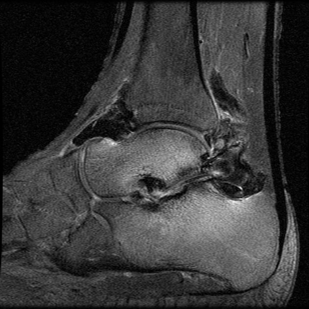
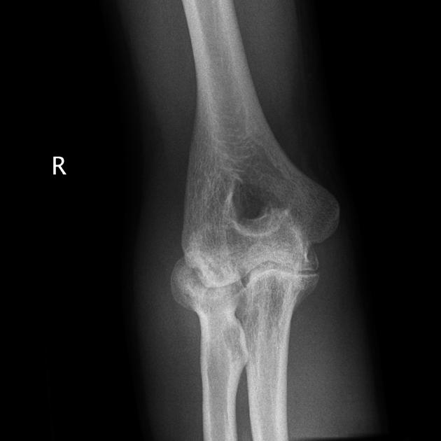
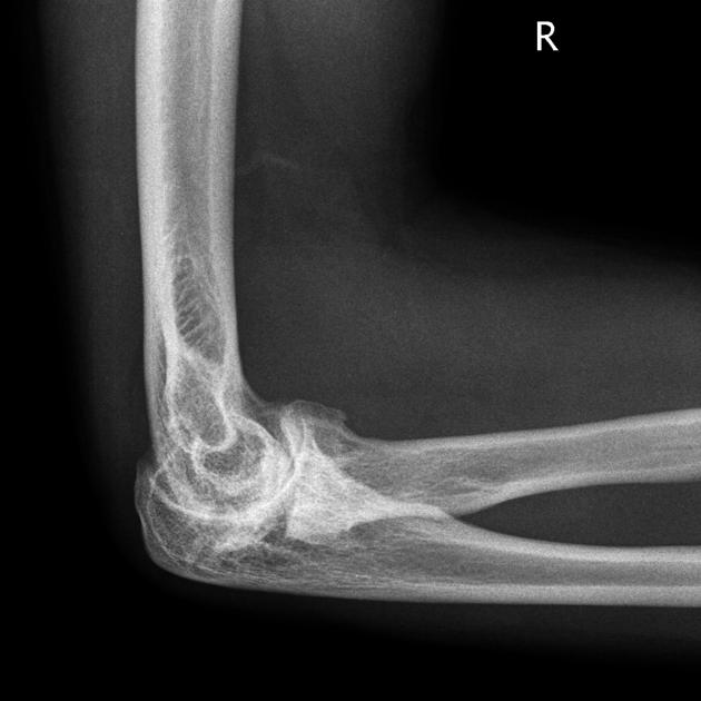
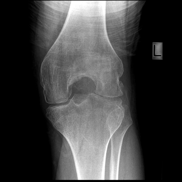
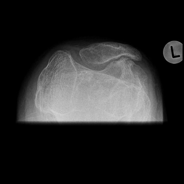
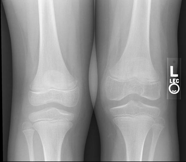
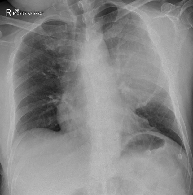
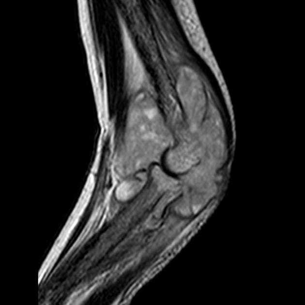
The hallmark of the disease is haemorrhage, particularly into joints and/or soft tissue, with several radiological consequences:
haemophilic arthropathy occurs in almost all individuals
haemophilic pseudotumour occurs in ~2%
soft tissue haematoma formation may lead to contractures 3
serious life-threatening haemorrhage (intracranial, thoracic, abdominal)
Treatment and prognosis
Treatment depends on the type, general severity, and current clinical state, and can be delivered episodically or prophylactically. Options for treatment include factor products (plasma-derived or recombinant) or novel medications such as emicizumab 10.
Prognosis depends on the severity and on the presence or absence of transfusion-related disease. Complications from HIV and cirrhosis are the leading causes of death. Life expectancy in those without HIV is ~62 years 2.
~15 times increased risk of death from intracranial haemorrhage (~1/3 of all deaths)
~50 times increased risk of death from non-intracranial haemorrhage


 Unable to process the form. Check for errors and try again.