
Haemorrhoids or anal cushions are normal vascular cushions of submucosal tissue present since birth in the anal canal and help in maintaining stool continence 2,6. Symptomatic pathological haemorrhoids occur secondary to raised intra-abdominal pressure 2.
On this page:
Epidemiology
The prevalence of haemorrhoids is ~20% (range 4-40%), and the actual prevalence remains unknown 2. It has an equal distribution between males and females 1. It is most commonly seen between 45 and 65 years of age and is more common in higher socioeconomic groups 1-3.
Associations
pregnancy 3
intra-abdominal masses 2
obesity 1
Diagnosis
The diagnosis is established by digital rectal examination and anoscope 1. Proctoscopy and colonoscopy are also considered in certain situations when the patient shows symptoms such as weight loss and bleeding 1,6.
Classification
Goligher’s classification is used in the grading of internal haemorrhoids 3:
grade 1: bleeding of haemorrhoids without prolapse
grade 2: prolapse of haemorrhoids on straining, which reduce spontaneously
grade 3: prolapse of haemorrhoids on straining, which require manual reduction
grade 4: prolapse is irreducible
Clinical presentation
Bleeding and pain associated with bowel movements is the most common presentation of haemorrhoids 1-3. The patient may also report a feeling of incomplete evacuation of bowel 2. Prolapse of haemorrhoids causes itching irritation, foreign body sensation and mucoid discharge 2.
Examination may reveal bright red blood, fissures, fistulas, skin tags, abscess and thrombosed or prolapsed anal cushions 1,3.
Pathology
Haemorrhoids are found in left lateral, right anterior and right posterior positions 2.
Symptomatic pathological haemorrhoids occur secondary to raised intra-abdominal pressure 2 and are classified into external and internal haemorrhoids based on their location relative to the dentate line 3. External haemorrhoids are located below the dentate line and are associated with thrombosis and pain. Internal haemorrhoids are situated above the dentate line and are associated with bleeding and prolapse 3.
Various pathological changes occur in the haemorrhoidal cushions in symptomatic haemorrhoids. These include 3
thrombosis
degeneration of collagen and elastic tissues
dilated and thin-walled vessels in submucosal plexus
loss of support from the Treitz muscle
abnormal vascular tone
activation of matrix metalloproteinase, vascular endothelial growth factor and transforming growth factor beta
Radiographic features
Although imaging modalities are not routinely used in the diagnosis of pathological haemorrhoids, it can aid in evaluation.
Ultrasound
Mosaic pattern, with turbulent flow in colour Doppler, is seen on ultrasound in grade 3 and grade 4 haemorrhoids 5.
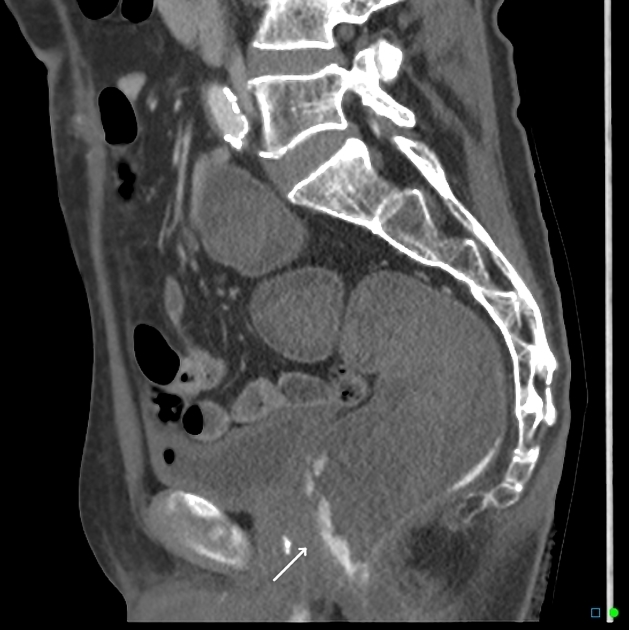
CT
Haemorrhoids and rectal varices appear similar and need to be carefully distinguished. Serpiginous veins are visible in haemorrhoids, which show poor or absent enhancement in the venous phase of contrast 4.
Treatment and prognosis
Conservative management includes increase in fibre and fluid intake and the use of stool softeners. Polythene glycol and docusate can be used for this purpose along with analgesics for pain relief 1. Minimally invasive techniques such as rubber band ligation, sclerotherapy, and infrared photocoagulation are also being routinely performed 1-3.
Surgical management includes haemorrhoidectomy, which can be performed as a daycare surgery. Haemorrhoidectomy can be either an open haemorrhoidectomy (Milligan-Morgan procedure) or closed haemorrhoidectomy (Ferguson procedure) 2.


 Unable to process the form. Check for errors and try again.