
The heart is a hollow, muscular organ of the middle mediastinum, designed to pump oxygenated blood around the systemic circulation and deoxygenated blood around the pulmonary circulation.
On this page:
Gross anatomy
The heart has a somewhat pyramidal form and is enclosed by the pericardium. Its base (roughly square-shaped) points posterior while its apex points to the left and inferiorly 7. It is positioned posteriorly to the body of the sternum, with one-third situated on the right and two-thirds on the left of the midline. Its left-sided orientation is formally known as laevocardia (cf. dextrocardia). The heart measures 12 x 8.5 x 6 cm and weighs ~310 g (males) and ~255 g (females) 1.
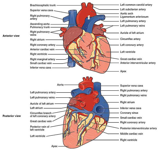
The heart is subdivided into chambers by septa into right and left halves, and a constriction subdivides each half of the organ into two cavities, the upper cavity being called the atrium and the lower the ventricle. The heart, therefore, consists of four chambers:
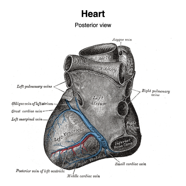
The division of the heart into four cavities is indicated on its surface by grooves. The atria are separated from the ventricles by the coronary sulcus (atrioventricular groove); this contains the trunks of the nutrient vessels of the heart and is deficient in front, where it is crossed by the root of the pulmonary artery. The interatrial groove, separating the two atria, is scarcely marked on the posterior surface while anteriorly it is hidden by the pulmonary trunk and ascending aorta.
The ventricles are separated by two grooves, one of which, the anterior longitudinal sulcus, is situated on the sternocostal surface of the heart, close to its left margin, the other posterior longitudinal sulcus, on the diaphragmatic surface near the right margin; these grooves extend from the base of the ventricular portion to a notch, the incisura apicis cordis, on the acute margin of the heart just to the right of the apex.
The cardiac wall consists of the following layers from inside to the outside:
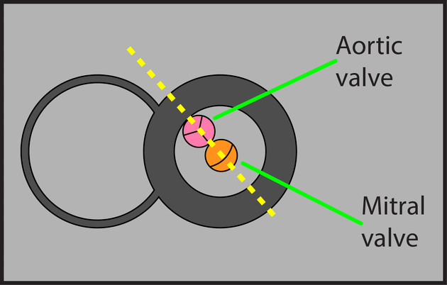
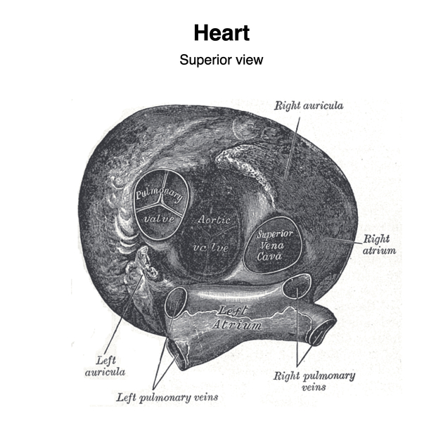
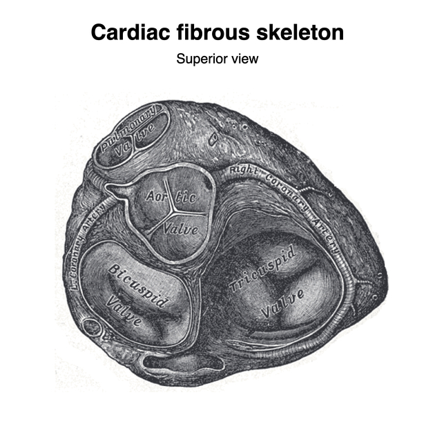
The outflow of each chamber is guarded by a heart valve:
-
atrioventricular valves between the atria and ventricles
mitral valve (bicuspid valve)
-
semilunar valves which are located in the outflow tracts of the ventricles
It is best to remember the four chambers and four valves in order of the series that blood travels through the heart:
venous blood returning from the body drains into the right atrium via the SVC, IVC and coronary sinus
the right atrium pumps blood through the tricuspid valve into the right ventricle
the right ventricle pumps blood through the pulmonary semilunar valve into the pulmonary trunk to be oxygenated in the lungs
blood returning from the lungs drains into the left atrium via the four pulmonary veins
the left atrium pumps blood through the bicuspid (mitral) valve into the left ventricle
the left ventricle pumps blood through the aortic semilunar valve into the ascending aorta to supply the body
The heart can be described as having the following surfaces:
-
posterior surface (base)
directed upward, backward and to the right
formed mainly by the left atrium 7 and little by the right atrium
-
apex
directed downward, forward and to the left
formed by the left ventricle 7
-
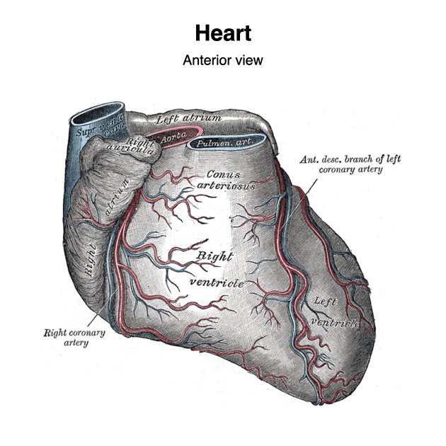
anterior (sternocostal) surface
directed forward, upward and to the left
formed mainly by the right ventricle inferiorly 7 and superiorly by the atria
-
inferior (diaphragmatic) surface
directed downward, slightly backward
formed by both ventricles 7
rests mainly upon the central tendon of the diaphragm
-
right surface
long; formed by right atrium superiorly 7 and right ventricle inferiorly
-
left (pulmonary) surface
shorter rounded; formed mainly by the left ventricle 7 and a little superiorly by the left atrium
Borders
The heart has four borders:
left border: left ventricle, left atrium, pulmonary trunk and arch of aorta
inferior border: right ventricle
superior border: right and left atria, SVC, ascending aorta and pulmonary trunk
See: Silhouette sign
Relations
anteriorly: the body of the sternum, and adjoining costal cartilages; left lung, and pleura (apex)
posteriorly: oesophagus, descending thoracic aorta, azygos, hemiazygos veins, and thoracic duct
superiorly: bifurcation of the main pulmonary trunk
inferiorly: diaphragm
laterally: lungs, pleura
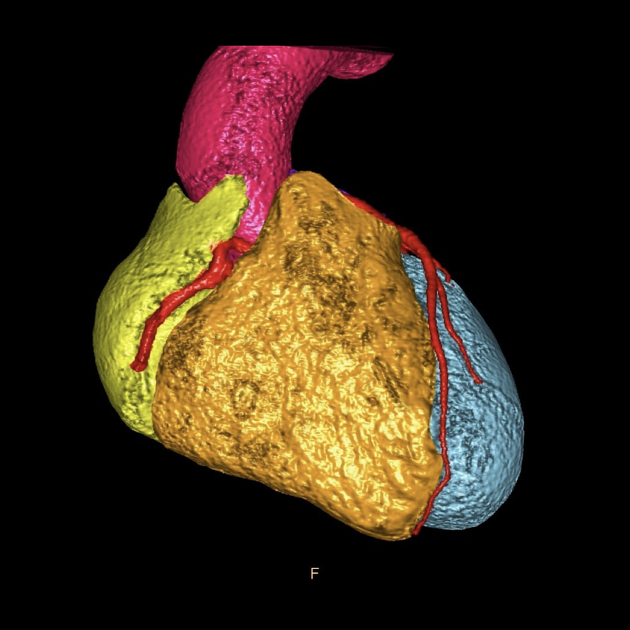
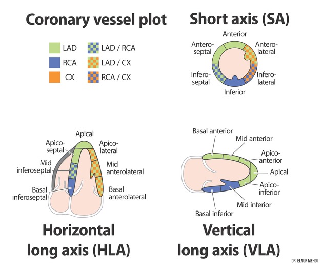
Arterial supply
Arterial supply is from the coronary arteries with coronary arterial dominance describing the dominant vessel supplying the interventricular septum. The vascular territories of the myocardium are divided into 17 myocardial segments according to the AHA nomenclature.
Venous drainage
Venous drainage is via the variable coronary veins and the coronary sinus.
Innervation
See main article: innervation of the heart.
Lymphatic drainage
Various lymphatic plexuses drain into a right cardiac collecting trunk (draining to anterior mediastinal nodes) and a left cardiac collecting trunk (draining to tracheobronchial nodes and onto paratracheal nodes).
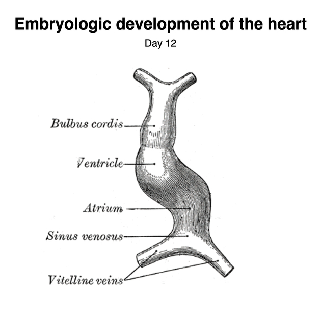
Embryological development
The heart develops from the fusion of two endocardial cardiac tubes of endodermal origin into a primitive heart tube which then undergoes a complex series of dilatations, twisting, and septation in the first month which is covered in detail in the article development of the heart.
Variant anatomy
The line can become somewhat blurred between what constitutes an anatomical variation and congenital heart disease but the key differentiator could be considered the presence or absence of symptoms in the majority of cases:
Eustachian valve: remnant valve of the IVC
Thebesian valve of the coronary sinus
There is also considerable variation in the anatomy of the coronary circulation and pulmonary veins.


 Unable to process the form. Check for errors and try again.