
Hepatic myelolipoma is a rare, benign fat-containing lesion of the liver, usually asymptomatic and found incidentally. Its diagnosis by imaging remains difficult because of a lack of pathognomonic signs. The definite diagnosis is by resection or biopsy.
On this page:
Epidemiology
Hepatic myelolipomas are extremely rare; only 17 cases have been described in the literature.
Clinical presentation
Hepatic myelolipomas are usually asymptomatic.
Pathology
These lipomatous tumours of the liver are extremely uncommon. Their composition is a mixture of fatty, muscular, haemopoietic, and vascular tissues and the histopathological spectrum includes angiomyolipomas, lipomas and myelolipomas.
The hepatic myelolipomas consist of adipose and myeloid tissue. Their pathogenesis, including hepatic localisation remains unclear, and an association with liver cell metaplasia has been suggested.
Radiographic features
Ultrasound
Appears as a hyperechoic well-circumscribed, lobulated mass.
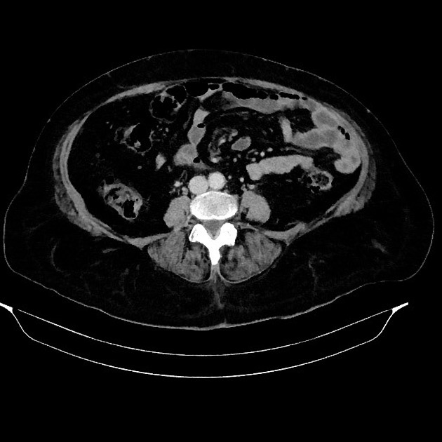
CT
Hypodense mass, with fatty attenuations ranging from -20 to -100 HU. Occasionally, the attenuation value is greater than zero, which is dependent on the relative proportions of fat and other elements in the tumour.
- unenhanced: lobulated well-circumscribed hypodense mass with fat density
- arterial phase: no enhancement
- portal venous phase: hypodense without enhancement
MRI
Appears as a well-defined mass with a pseudocapsule, heterogeneous fatty architecture and contents.
Treatment and prognosis
Surgical treatment is reserved for symptomatic patients or in case of diagnostic uncertainty.
Malignant transformation has not been described.
History and etymology
The first case of a hepatic myelolipoma was described in France by Grosdidier in 1973.
Differential diagnosis
- hepatic angiomyolipoma
- hepatic lipoma
- hepatic adenoma
- haemangioma
- hepatic adrenal rest tumour
- focal nodular hyperplasia
For a wider range of differentials, read more in our article on fat containing liver lesions.


 Unable to process the form. Check for errors and try again.