Citation, DOI, disclosures and article data
Citation:
St-Amant M, Sharma R, Saber M, et al. Hippocampal sulcus remnant cyst. Reference article, Radiopaedia.org (Accessed on 28 Mar 2025) https://doi.org/10.53347/rID-24971
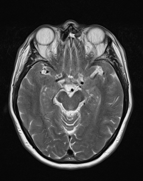
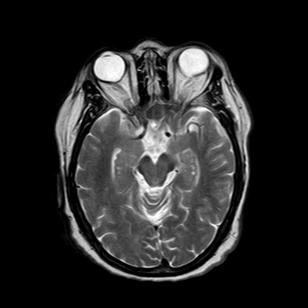
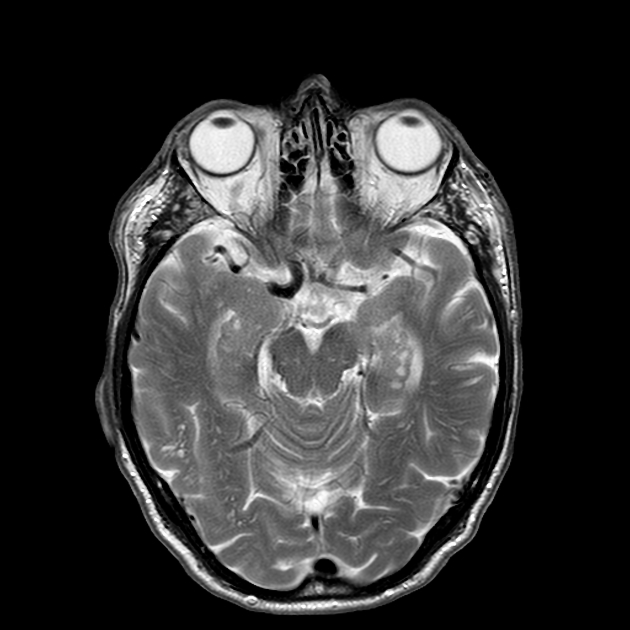
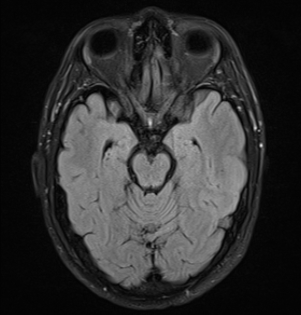
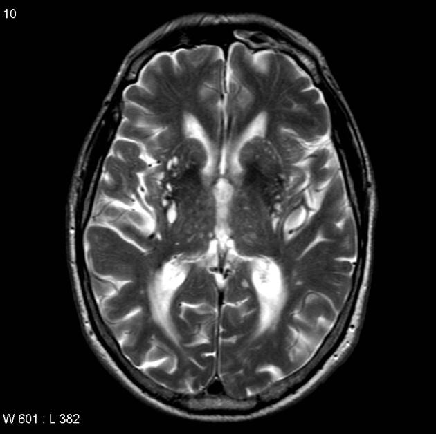
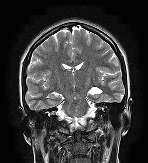
Hippocampal sulcus remnant cysts, also known as hippocampal cavities, are remnants of incomplete involution of the embryonic hippocampal fissure and are an incidental finding.
They are seen in ~25% (range 10-40%) of the adult population 1,3.
These are asymptomatic and benign entities, not associated with seizures or epilepsy 5.
MRI
They consist of small (1-2 mm) cystic spaces that follow CSF on all sequences. They are located between the dentate gyrus and the cornu ammonis, at the lateral aspect of the hippocampal region, near the apex of the hippocampal fold 4.
-
1. Sasaki M, Sone M, Ehara S et-al. Hippocampal sulcus remnant: potential cause of change in signal intensity in the hippocampus. Radiology. 1993;188 (3): 743-6. Pubmed citation
-
2. Li Y, Li J, Segal S et-al. Hippocampal cerebrospinal fluid spaces on MR imaging: Relationship to aging and Alzheimer disease. AJNR Am J Neuroradiol. 2006;27 (4): 912-8. AJNR Am J Neuroradiol (full text) - Pubmed citation
-
3. Atlas SW. Magnetic Resonance Imaging Of The Brain And Spine. Lippincott Williams & Wilkins. (2009) ISBN:078176985X. Read it at Google Books - Find it at Amazon
-
4. Bastos-Leite AJ, van Waesberghe JH, Oen AL, van der Flier WM, Scheltens P, Barkhof F. Hippocampal sulcus width and cavities: comparison between patients with Alzheimer disease and nondemented elderly subjects. (2006) AJNR. American journal of neuroradiology. 27 (10): 2141-5. Pubmed
-
5. Hassankhani A, Stein J, Haboosheh A, Vossough A, Loevner L, Nabavizadeh S. Anatomical Variations, Mimics, and Pitfalls in Imaging of Patients with Epilepsy. J Neuroimaging. 2020;31(1):20-34. doi:10.1111/jon.12809 - Pubmed
Promoted articles (advertising)


 Unable to process the form. Check for errors and try again.