
Interventricular foramen (of Monro)
Citation, DOI, disclosures and article data
At the time the article was created Jeremy Jones had no recorded disclosures.
View Jeremy Jones's current disclosuresAt the time the article was last revised Craig Hacking had the following disclosures:
- Philips Australia, Paid speaker at Philips Spectral CT events (ongoing)
These were assessed during peer review and were determined to not be relevant to the changes that were made.
View Craig Hacking's current disclosures- Interventricular foramen
- Foramen of Monro
- Interventricular foramina
- Foramina of Monro
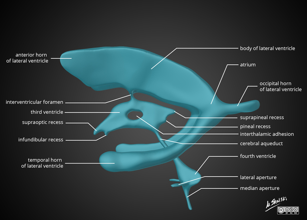
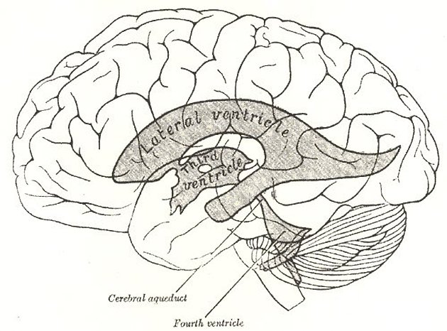
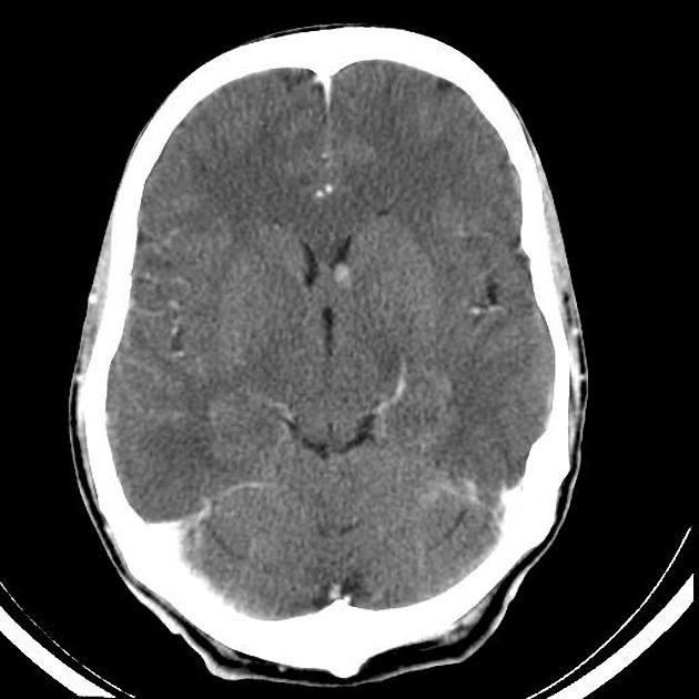
The interventricular foramen, also known as foramen of Monro, is part of the ventricular system and the connection between the third ventricle and the lateral ventricle.
These paired foramina allow for the flow of cerebrospinal fluid between lateral ventricles and third ventricles, and effacement or blockage results in non-communicating obstructive hydrocephalus.
On this page:
Gross anatomy
Each foramen of Monro lies between the roof and anterior wall of the third ventricle behind the column and body of the fornix and anterior to the thalamus 4.
Several structures pass through each foramen (in addition to CSF) 4:
arteries: distal branches of the medial posterior choroidal arteries
veins: thalamostriate, superior choroidal and septal
If the ventricles are small, then each foramen is a small crescent, concave anteriorly (indented by the fornix). On the other hand, if the ventricles are large, each foramen becomes more circular in cross-section 4.
History and etymology
The structure was named after the Scottish physician Alexander Monro (1733-1817), who first described it in 1783 3. It should be noted, however, that in his description he acknowledges that these communications were known about and previously described as far back as Galen, and this is another example of Stigler's law of eponymy 4.
To be precise, it should also be noted that the individual who described the foramen is Alexander Monro secundus (the second). His father and his son were both also called Alexander Monro (primus and tertius respectively) and all three of them held the chair of anatomy at the University of Edinburgh 4.
Related pathology
References
- 1. Stranding S. Gray's anatomy. Churchill Livingstone. (2005) ISBN:0443071683. Read it at Google Books - Find it at Amazon
- 2. Ross LMMP. Atlas of anatomy. George Thieme Verlag. (2007) ISBN:3131421215. Read it at Google Books - Find it at Amazon
- 3. Clarke E. The Human Brain and Spinal Cord. Norman Publishing. (1996) ISBN:0930405250. Read it at Google Books - Find it at Amazon
- 4. Tubbs RS, Oakes P, Maran IS, Salib C, Loukas M. The foramen of Monro: a review of its anatomy, history, pathology, and surgery. (2014) Child's nervous system : ChNS : official journal of the International Society for Pediatric Neurosurgery. 30 (10): 1645-9. doi:10.1007/s00381-014-2512-6 - Pubmed
Incoming Links
- Medial posterior choroidal artery
- Central neurocytoma
- Germinal matrix haemorrhage
- Cerebrospinal fluid
- Lateral ventricle
- Ventricular system
- Hydrocephalus
- Stigler's law of eponymy
- Connatal cyst
- Head ultrasound
- Hippocampal commissure
- Megalencephaly
- Internal cerebral vein
- Transsphenoidal hypophysectomy
- Deep cerebral veins
- Caudothalamic groove
- Third ventricle
- Transcranial Doppler sonography (ultrasound)
- Thalamus
- Cavum vergae cyst
- Foramen of Monro obstruction
- Colloid cyst with hydrocephalus
- Colloid cyst
- Right frontal external ventricular drain
- Colloid cyst of the third ventricle
- Coagulopathy related intracerebral hemorrhage on background of uremic encephalopathy
- Bilateral stenosis of the foramina of Monro
- Central neurocytoma
- Bilateral foramen of Monro stenosis
- Cytotoxic lesion of the corpus callosum
- Adamantinomatous craniopharyngioma
- Tuberous sclerosis with giant cell astrocytoma
- Colloid cyst
- Colloid cyst
- Colloid cyst of the 3rd ventricle
- Tuberous sclerosis
- Colloid cyst
- Central neurocytoma
- Colloid cyst
- Colloid cyst of the third ventricle
Related articles: Anatomy: Brain
-
brain
- grey matter
- white matter
-
cerebrum
-
cerebral hemisphere (telencephalon)
- cerebral lobes and gyri
- frontal lobe
- parietal lobe
-
occipital lobe
- occipital pole
- lingual gyrus
- fusiform gyrus (Brodmann area 37)
- calcarine (visual) cortex
- cuneus
- temporal lobe
- basal forebrain
- limbic system
- insula
-
cerebral sulci and fissures (A-Z)
- calcarine fissure
- callosal sulcus
- central (Rolandic) sulcus
- cingulate sulcus
- collateral sulcus
- inferior frontal sulcus
- inferior occipital sulcus
- inferior temporal sulcus
- interhemispheric fissure
- intraparietal sulcus
- lateral (Sylvian) sulcus
- lateral occipital sulcus
- marginal sulcus
- occipitotemporal sulcus
- olfactory sulcus
- paracentral sulcus
- paraolfactory sulcus
- parieto-occipital fissure
- posterior parolfactory sulcus
- precentral sulcus
- preoccipital notch
- postcentral sulcus
- rhinal sulcus
- rostral sulcus
- subparietal sulcus
- superior frontal sulcus
- superior occipital sulcus
- superior temporal sulcus
- cortical histology
- cerebral lobes and gyri
- white matter tracts
- deep grey matter
-
pituitary gland
- posterior pituitary and stalk (part of diencephalon)
- anterior pituitary
- inferior hypophyseal arterial circle
- diencephalon
-
cerebral hemisphere (telencephalon)
-
brainstem
- midbrain (mesencephalon)
- pons (part of metencephalon)
- medulla oblongata (myelencephalon)
- white matter
-
grey matter
- non-cranial nerve
-
cranial nerve nuclei
- oculomotor nucleus
- Edinger-Westphal nucleus
- trochlear nucleus
- motor nucleus of CN V
- mesencephalic nucleus of CN V
- main sensory nucleus of CN V
- spinal nucleus of CN V
- abducent nucleus
- facial nucleus
- superior salivatory nucleus
- cochlear nuclei
- vestibular nuclei
- inferior salivatory nucleus
- solitary tract nucleus
- ambiguus nucleus
- dorsal vagal motor nucleus
- hypoglossal nucleus
-
cerebellum (part of metencephalon)
- vermis
- cerebellar hemisphere
- cerebellar peduncles
- cranial meninges (meninx primitiva)
- CSF spaces
-
cranial nerves (mnemonic)
- olfactory nerve (CN I)
- optic nerve (CN II)
- oculomotor nerve (CN III)
- trochlear nerve (CN IV)
- trigeminal nerve (CN V) (mnemonic)
- abducens nerve (CN VI)
- facial nerve (CN VII) (segments mnemonic | branches mnemonic)
-
vestibulocochlear nerve (CN VIII)
- vestibular ganglion (Scarpa's ganglion)
- glossopharyngeal nerve (CN IX)
- vagus nerve (CN X)
- spinal accessory nerve (CN XI)
- hypoglossal nerve (CN XII)
- functional neuroanatomy
- CNS development
- cerebral vascular supply
- arteries
- vascular territories
-
circle of Willis
- internal carotid artery (ICA) (segments)
- vertebral artery
-
normal variants
- intracranial arterial fenestration
- internal carotid artery (ICA)
- anterior cerebral artery (ACA)
- middle cerebral artery (MCA)
- posterior cerebral artery (PCA)
- basilar artery
- persistent carotid-vertebrobasilar artery anastomoses (mnemonic)
- vertebral artery
- ophthalmic artery
-
cerebral venous system
-
dural venous sinuses
- basilar venous plexus
- cavernous sinus (mnemonic)
- clival diploic veins
- inferior petro-occipital vein
- inferior petrosal sinus
- inferior sagittal sinus
- intercavernous sinus
- internal carotid artery venous plexus of Rektorzik
- jugular bulb
- marginal sinus
- occipital sinus
- sigmoid sinus
- sphenoparietal sinus
- straight sinus
- superior petrosal sinus
- superior sagittal sinus
- torcula herophili
- transverse sinus
-
cerebral veins
-
superficial veins of the brain
- superior cerebral veins (superficial cerebral veins)
- inferior cerebral veins
- superficial middle cerebral vein
- superior anastomotic vein (of Trolard)
- inferior anastomotic vein (of Labbe)
-
superficial veins of the brain
-
deep veins of the brain
- great cerebral vein (of Galen)
- venous circle of Trolard
- normal variants
-
dural venous sinuses
- arteries
- glymphatic pathway


 Unable to process the form. Check for errors and try again.