Citation, DOI, disclosures and article data
Citation:
Weerakkody Y, Campos A, Feger J, et al. Iron overload cardiomyopathy. Reference article, Radiopaedia.org (Accessed on 23 Mar 2025) https://doi.org/10.53347/rID-32513
Iron overload cardiomyopathy (IOC) refers to a secondary form of cardiomyopathy resulting from the accumulation of iron in the myocardium. It occurs mainly due to genetically determined disorders of iron metabolism (e.g. cardiomyopathy in hemochromatosis, thalassemia 6,7) or multiple transfusions.
Pathology
It been mostly described as a dilated cardiomyopathy, characterized by:
In primary hemochromatosis leading to iron overload, the cardiomyopathy is classically categorized as an infiltrative cause of restrictive cardiomyopathy. While in those with secondary hemochromatosis, there may be severe diastolic LV dysfunction in the early stages of the disease, before LVEF is affected 3.
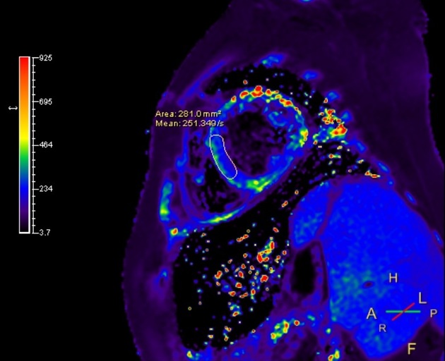
Radiographic features
MRI
CMR-derived T2* relaxation time is the mainstay for the quantitative assessment of cardiac iron deposition.
Measured in a full-thickness area of interest in the interventricular septum, T2* is highly representative of global myocardial iron.
A value of 20 ms is considered to be the threshold for myocardial siderosis.
-
1. Gujja P, Rosing D, Tripodi D, Shizukuda Y. Iron Overload Cardiomyopathy: Better Understanding of an Increasing Disorder. J Am Coll Cardiol. 2010;56(13):1001-12. doi:10.1016/j.jacc.2010.03.083 - Pubmed
-
2. Murphy CJ, Oudit GY. Iron-overload cardiomyopathy: pathophysiology, diagnosis, and treatment. J. Card. Fail. 2010;16 (11): 888-900. doi:10.1016/j.cardfail.2010.05.009 - Pubmed citation
-
3. Kremastinos DT, Farmakis D. Iron overload cardiomyopathy in clinical practice. Circulation. 2011;124 (20): 2253-63. doi:10.1161/CIRCULATIONAHA.111.050773 - Pubmed citation
-
4. Kobayashi T, Tadokoro H, Matsumoto K. An autopsy case of secondary iron-overload cardiomyopathy. Intern. Med. 2014;52 (12): 1369-73. Pubmed citation
-
5. Shamsian B, Esfahani S, Milani H et al. Magnetic Resonance Imaging in the Evaluation of Iron Overload: A Comparison of MRI, Echocardiography and Serum Ferritin Level in Patients with β-Thalassemia Major. Clin Imaging. 2012;36(5):483-8. doi:10.1016/j.clinimag.2011.11.029 - Pubmed
-
6. Anderson LJ, Westwood MA, Prescott E et-al. Development of thalassaemic iron overload cardiomyopathy despite low liver iron levels and meticulous compliance to desferrioxamine. Acta Haematol. 2006;115 (1-2): 106-8. doi:10.1159/000089475 - Pubmed citation
-
7. Pennell D. MRI and Iron-Overload Cardiomyopathy in Thalassaemia. Circulation. 2006;113(11):f43-4. - Pubmed
Promoted articles (advertising)


 Unable to process the form. Check for errors and try again.