
Knee bursae
Citation, DOI, disclosures and article data
Citation:
Su S, Murphy A, Feger J, et al. Knee bursae. Reference article, Radiopaedia.org (Accessed on 22 Mar 2025) https://doi.org/10.53347/rID-32169
rID:
32169
Article created:
17 Nov 2014,
Shu Su
Disclosures:
At the time the article was created Shu Su had no recorded disclosures.
View Shu Su's current disclosures
Last revised:
Disclosures:
At the time the article was last revised Andrew Murphy had no recorded disclosures.
View Andrew Murphy's current disclosures
Revisions:
17 times, by
12 contributors -
see full revision history and disclosures
Systems:
Sections:
Synonyms:
- Bursae of the knee
- Bursal spaces around the knee
- Bursal spaces of the knee
Knee bursae are sacs surrounding the knee joint that are filled with synovial fluid. They facilitate movement and reduce friction where tendons or muscles pass over bony prominences. The knee bursae can be either communicating or non-communicating with the knee joint itself.
On this page:
Article:
Images:
Images:
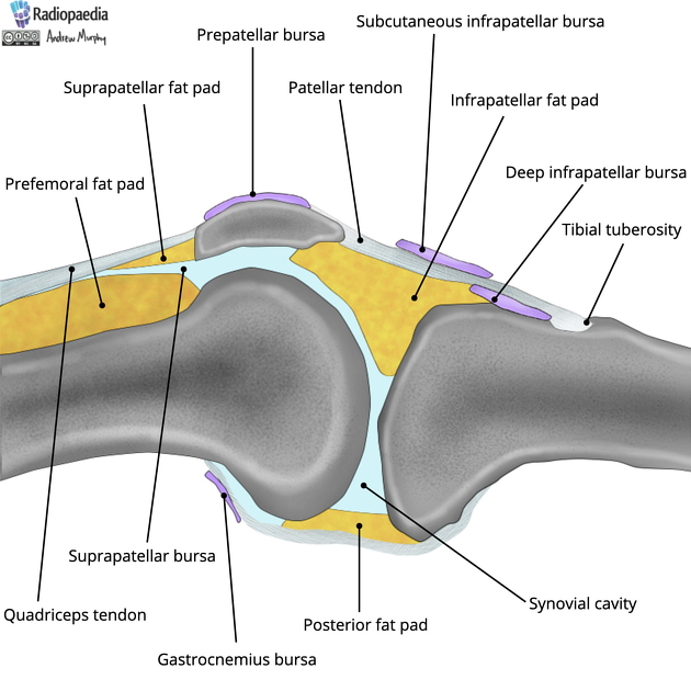
Gross anatomy
There are four bursae anterior to the knee joint:
- suprapatellar bursa: located between the femur and quadriceps femoris, it is attached to the articularis genu muscle and usually communicates with the synovial cavity
- subcutaneous prepatellar bursa: between the skin and patella
- subcutaneous infrapatellar bursa: between the skin and tibial tuberosity
- deep infrapatellar bursa: between patella ligament and upper tibia
Two bursae are located laterally:
- fibular collateral ligament-biceps femoris bursa: between the lateral collateral ligament and the biceps femoris tendon
- between the lateral collateral ligament and the capsule where it overlies the popliteus muscle
Two bursae are located medially:
- between the medial collateral ligament and the pes anserinus
- between the medial collateral ligament and the capsule, tibia and semimembranosus tendon
There are four bursae posterior to the knee joint:
- between the capsule and medial head of gastrocnemius; communicates with the synovial cavity
- semimembranosus bursa: between semimembranosus and the medial head of gastrocnemius; may communicate with the bursa under the medial head of the gastrocnemius and thereby the synovial cavity
- lateral gastrocnemius bursa: between the capsule and lateral head of gastrocnemius; may communicate with the synovial cavity in some people
- popliteus bursa: between popliteus tendon and posterior tibia and fibula; communicates with the synovial cavity 1
Related pathology
See also
References
- 1. Sinnatamby CS. Last's Anatomy. Elsevier Health Sciences. (2011) ISBN:0702048399. Read it at Google Books - Find it at Amazon
- 2. Hirji Z, Hunjun JS, Choudur HN. Imaging of the bursae. (2011) Journal of clinical imaging science. 1: 22. doi:10.4103/2156-7514.80374 - Pubmed
Incoming Links
Articles:
Related articles: Anatomy: Lower limb
- skeleton of the lower limb
- joints of the lower limb
-
hip joint
- ligaments
- muscles
- additional structures
- hip joint capsule
- zona orbicularis
- iliotibial band
-
hip bursae
- anterior
- iliopsoas bursa (iliopectineal bursa)
- lateral
- subgluteal bursae
- greater trochanteric bursa (subgluteus maximus bursa)
- subgluteus medius bursa
- subgluteus minimus bursa
- gluteofemoral bursa
- subgluteal bursae
- postero-inferior
- anterior
- ossification centers
-
knee joint
- ligaments
- anterior cruciate ligament
- posterior cruciate ligament
- medial collateral ligament
- lateral collateral ligament
- meniscofemoral ligament (mnemonic)
-
posterolateral ligamentous complex
- arcuate ligament
- patellar tendon and quadriceps tendon
- anterolateral ligament
- posterior oblique ligament
- oblique popliteal ligament
- medial patellofemoral ligament
- additional structures
- extensor mechanism of the knee
- groove for the popliteus tendon
- knee bursae
- anterior bursae
- medial bursae
- lateral bursae
- posterior bursae
- knee capsule
- lateral patellar retinaculum
- medial patellar retinaculum
- menisci
- pes anserinus (mnemonic)
- ossification centers
- ligaments
- tibiofibular joints
-
ankle joint
- regional anatomy
- medial ankle
- lateral ankle
- anterior ankle
- ligaments
- medial collateral (deltoid) ligament
- lateral collateral ligament
- additional structures
- ankle bursae
- ossification centers of the ankle
- variants
- regional anatomy
- foot joints
- subtalar joint
- mid-tarsal (Chopart) joint
-
tarsometatarsal (Lisfranc) joint
- ligaments
- intermetatarsal joint
- metatarsophalangeal joint
- interphalangeal joint
- ossification centers
-
hip joint
- spaces of the lower limb
-
muscles of the lower limb
- muscles of the pelvic group
- muscles of the thigh
- muscles of the leg
- anterior compartment of the leg
- posterior compartments of the leg
- lateral compartment of the leg
- muscles of the foot
- dorsal muscles
- plantar muscles
- 1st layer
- 2nd layer
- 3rd layer
- 4th layer
- accessory muscles of the lower limb
- accessory gluteal muscles
-
accessory muscles of the ankle
- accessory peroneal muscles
- accessory flexor digitorum longus muscle
- accessory soleus muscle
- peroneocalcaneus internus muscle
- tibiocalcaneus internus muscle
- extensor hallucis capsularis tendon
- anterior fibulocalcaneus muscle
- accessory extensor digiti secundus muscle
- tibioastragalus anticus of Gruber muscle
- vascular supply of the lower limb
- arterial supply of the lower limb
- venous drainage of the lower limb
- innervation of the lower limb
- lymphatic system of the lower limb
- lymphatic pathways
- anteromedial group
- anterolateral group
- posteromedial group
- posterolateral group
- lower limb lymph nodes
- lymphatic pathways


 Unable to process the form. Check for errors and try again.