
Knuckle pads, also known as Garrod pads, Garrod nodes or holoderma, are benign, subcutaneous, fibrofatty growths that overlie the interphalangeal joints of the fingers or toes 1,2. These are a rare form of superficial musculoskeletal fibromatoses.
On this page:
Clinical presentation
Knuckle pads present as bilaterally asymmetrical subcutaneous growths that overlie the dorsal aspect of the finger or toe interphalangeal joint or, less commonly, the metacarpophalangeal joint 2. Knuckle pads are usually skin-coloured, firm and painless. However, they can lead to discomfort and large knuckle pads can cause difficulty moving the fingers or toes 3.
Epidemiology
Knuckle pads can be genetic and occur more regularly in people with another type of fibromatosis, such as Dupuytren contracture or Peyronie disease 3. Patients most commonly present with knuckle pads as adults, after the age of 30 years 1.
Pathology
Knuckle pads are a type of superficial musculoskeletal fibromatoses. They are subcutaneous nodules overlying the dorsal aspect of the extensor tendon at the level of the interphalangeal or metacarpophalangeal joint.
Aetiology
The aetiology is unknown and is usually considered idiopathic but there are associated with repetitive trauma (e.g. boxing, swimming, carpet laying), and other fibromatoses (e.g. Duputren disease, Peyronine disease, Ledderhose disease) 2,3,5.
Microscopic appearance
Focal proliferation of myofibroblasts and decrease in elastic filaments in the deep, reticular dermis 2.
Radiographic features
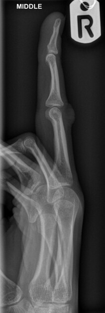
Plain radiograph
Soft tissue swelling overlying the dorsal aspect of the interphalangeal joint.
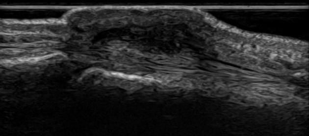
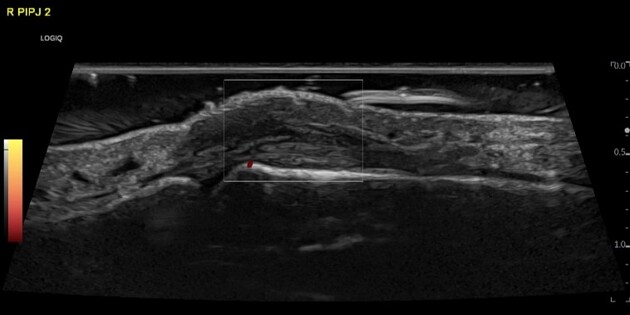
Ultrasound
On B-mode, they are seen as subcutaneous hypoechoic nodules with irregular borders. Doppler features include 2:
absence of internal flow signals on colour or power Doppler
peripheral hypervascularisation (rarely)
normal appearance of the adjacent joints and tendons is expected
MRI
T1: low signal
T2: intermediate signal
T2FS: high signal 4
Differential diagnoses
tenosynovial giant cell tumour 2,4
subcutaneous nodules, e.g. rheumatoid nodules, Herbeden's nodes, Bouchard's nodes, gouty tophi 2,4
tenosynovitis/synovitis, particularly if multifocal 2,5


 Unable to process the form. Check for errors and try again.