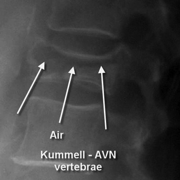
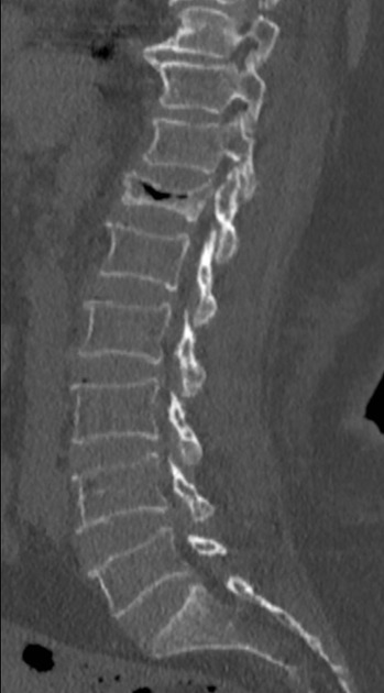
Kümmell disease represents delayed (usually two weeks) vertebral body collapse due to ischaemia and non-union of anterior vertebral body wedge fractures after minor trauma.
Radiographic features
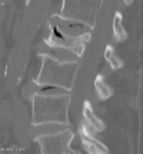
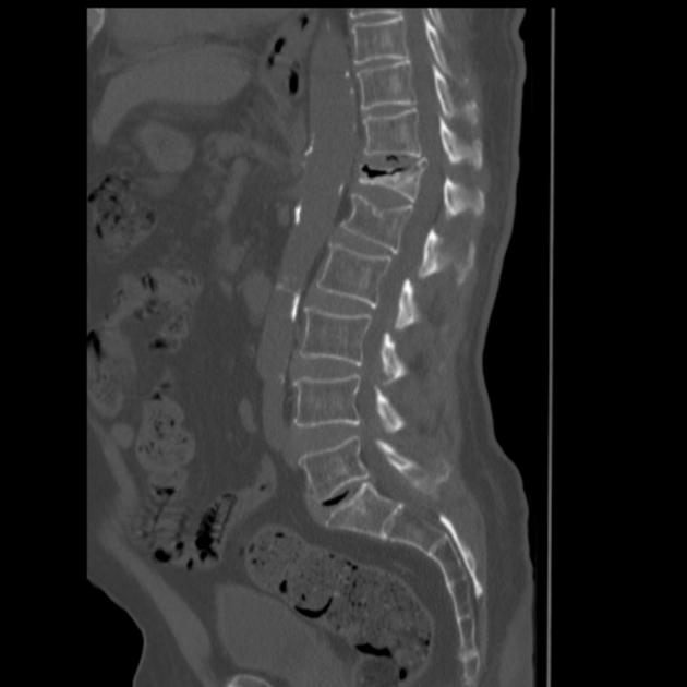
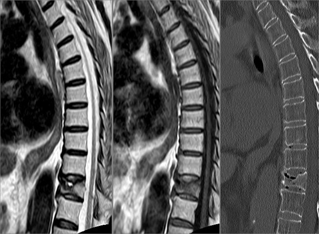
Plain radiograph / CT
shows collapse of affected vertebrae (typically lower thoracic and upper lumbar)
an intravertebral vacuum cleft and fluid may be seen (accentuated on extension stress lateral views), although this is non-specific
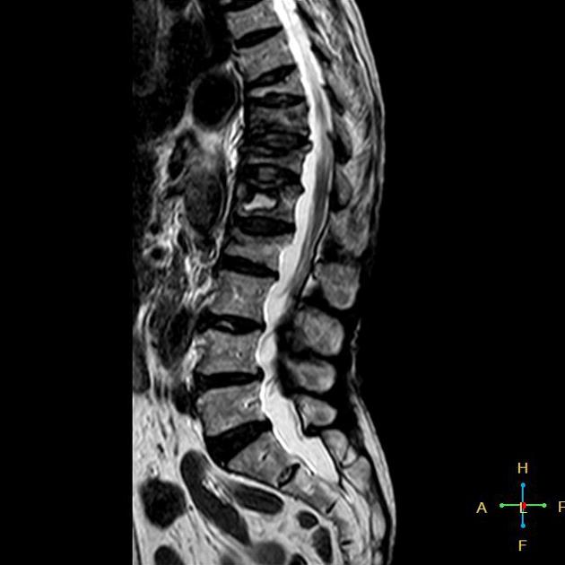
MRI
the intravertebral vacuum cleft is usually seen as low signal intensity with all sequences
if intravertebral fluid is present it is low T1 signal and high T2 signal
The severity of the vertebral collapse has been found to be higher in those who have intravertebral gas compared to those who only have intravertebral fluid, signifying that the presence of intravertebral gas is possibly a more advanced stage of the disease 1.
History and etymology
This condition was first described by the German surgeon Hermann Kümmell (1852-1937) in 1891 4,8.
1. Yu C, Hsu C, Shih T, Chen B, Fu C. Vertebral Osteonecrosis: MR Imaging Findings and Related Changes on Adjacent Levels. AJNR Am J Neuroradiol. 2007;28(1):42-7. PMC8134120 - Pubmed
2. Baur A, Stäbler A, Arbogast S, Duerr H, Bartl R, Reiser M. Acute Osteoporotic and Neoplastic Vertebral Compression Fractures: Fluid Sign at MR Imaging. Radiology. 2002;225(3):730-5. doi:10.1148/radiol.2253011413 - Pubmed
3. Freedman B & Heller J. Kummel Disease: A Not-So-Rare Complication of Osteoporotic Vertebral Compression Fractures. J Am Board Fam Med. 2009;22(1):75-8. doi:10.3122/jabfm.2009.01.080100 - Pubmed


 Unable to process the form. Check for errors and try again.