The left superior intercostal vein drains the left posterosuperior hemithorax and is considered to be part of the azygos venous system even though it does not directly drain into the azygos vein. It should not be confused for the supreme intercostal vein, which drains the 1st intercostal space.
On this page:
Gross anatomy
Origin
The left superior intercostal vein forms by the union of the 2nd to 4th left posterior intercostal veins.
Course
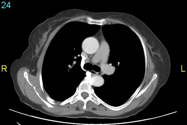
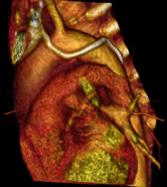
It courses superiorly to the left of the midline before arching lateral to the distal aortic arch to drain into the left brachiocephalic vein anteriorly. It courses medial to phrenic nerve and lateral to vagus nerve.
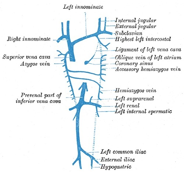
It typically communicates with the accessory hemiazygos vein inferiorly.
Tributaries
left 2nd to 4th posterior intercostal veins
Variant anatomy
may not communicate with the accessory hemiazygos (25%) 1
left azygos (or hemiazygos) lobe may be caused by an aberrant left superior intercostal vein (analogous to the azygos lobe) 1
hemiazygos and accessory hemiazygos veins may drain directly into the left brachiocephalic vein via the left superior intercostal vein 1
may be enlarged in the presence of congenital azygos, hemiazygos or accessory hemiazygos vein absence (rare)
Radiographic appearance
Plain film
-
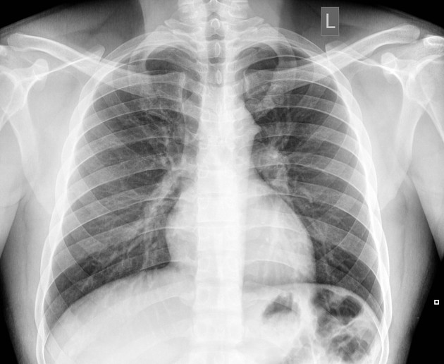
on ~5% of frontal chest x-rays, it may form an "aortic nipple" next to the aortic arch as it is imaged perpendicular to the x-ray beam 1,2
this should measure less than 4.5 mm and if larger it may indicate underlying pathology (e.g. SVC obstruction or azygos continuation of the IVC) 2


