
Musculoskeletal involvement of leukaemia is not always apparent on imaging, although the disease is per se characterised by leukaemic infiltration of bone marrow.
Leukaemia is a haematological neoplasm characterised by the overproduction of immature (blasts) or abnormally differentiated cells of the haematopoietic system in the bone marrow that often, but not always, extends into the peripheral blood and, therefore, may involve multiple organs, including also the muscles and soft tissues.
Radiographic features
-
manifestations of bone marrow replacement
although most plain radiographs appear normal, osteopenia is the most frequent finding 1,2
-
radiolucent metaphyseal bands
paediatric patients, up to 40% of those with ALL 2
they represent the osteopenia in the most fast-growing parts of long bones 2
alternating radiolucent and radiodense metaphyseal lines are classically described in treated patients
-
coarse trabeculation
increased number of trabeculae can lead to an osteosclerotic appearance 2, which is commonly seen as a result of secondary myelofibrosis
vertebral body collapse (vertebra plana)
-
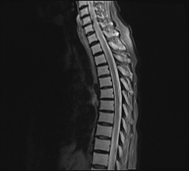
MRI
T1: diffuse low signal
T2: increased signal
T1 C+ (Gd): diffuse enhancement
-
bone involvement without marrow involvement
can be seen due to direct invasion or haematogenous seeding 2
bone lytic lesions
-
intramuscular leukaemic infiltrates
rare 1
focal soft tissue deposits forming mass-like lesion within the muscles
usually demonstrates contrast enhancement
iso signal compared to the background muscle on T1 and increased signal on T2 weighted images 1
Skin involvement, known as leukaemia cutis, represents focal deposits of leukaemic cells in the epidermis, dermis, or subcutaneous tissues 2
See also
-
systemic involvement of leukaemia


 Unable to process the form. Check for errors and try again.