
Mandible
Updates to Article Attributes
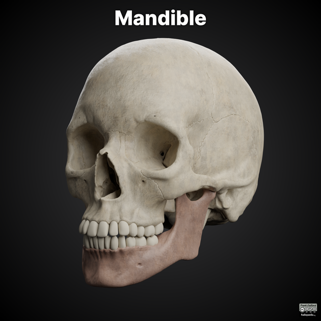
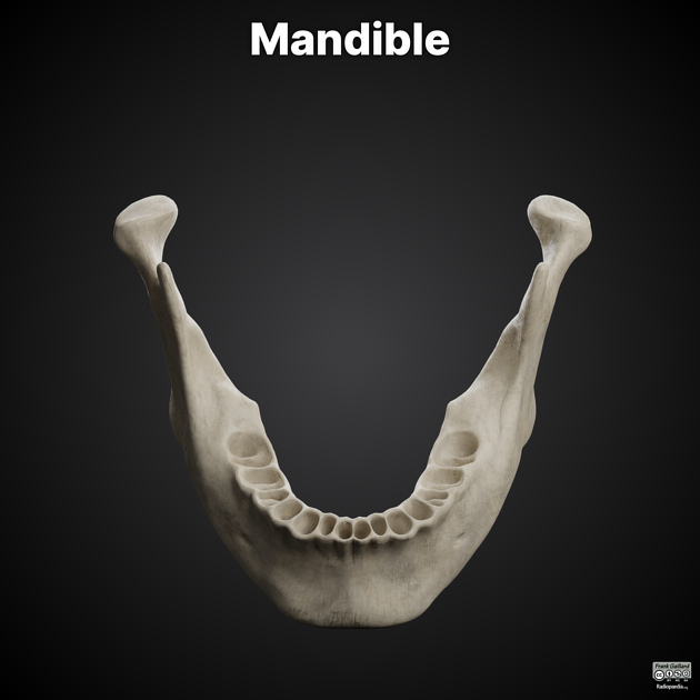
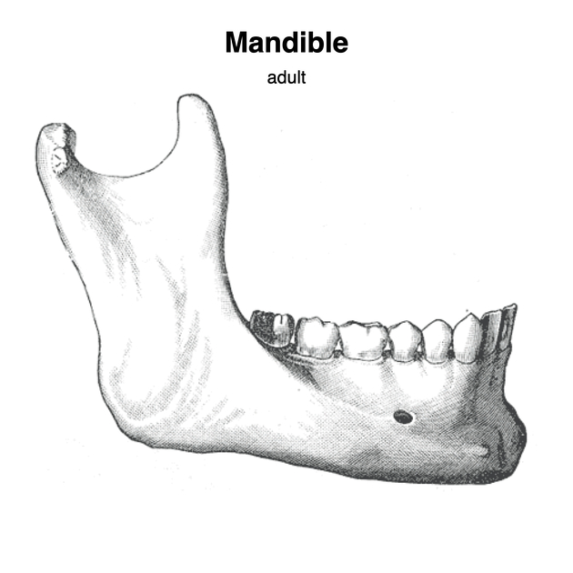
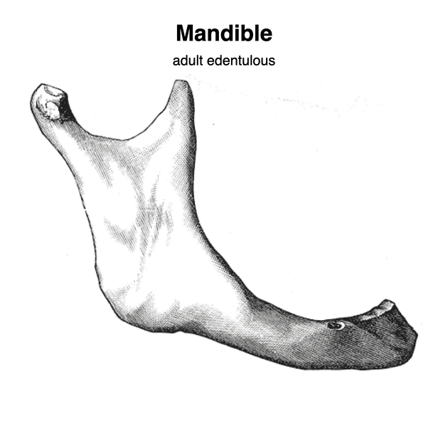
The mandible consists of a curved, horizontal portion, the body, and two perpendicular portions, the rami, which unite with the ends of the body nearly at right angles (angle of the jaw). It articulates with both temporal bones at the mandibular fossa at the temporomandibular joints (TMJ).
Osteology
Body
The body of the mandible is curved, somewhat like a horseshoe, and two surface and two borders:
- external surface
- midline ridge indicating the symphysis
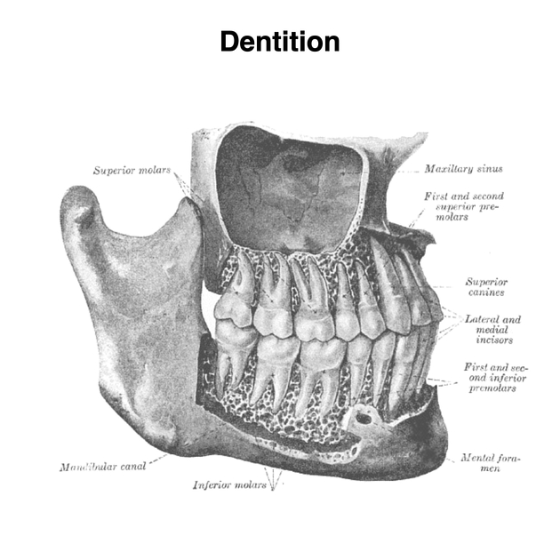
- mental foramen: inferior to second premolar tooth, midway between the superior and inferior borders; allows for the passage of the mental vessels and mental nerve
- internal surface
- concave from side-to-side
- origin of geniohyoid (from mental spines) and geniohyoid (median ridge or impression)
- fossae for the submental and submandibular salivary glands
- superior (or alveolar) border
- wider behind than in front
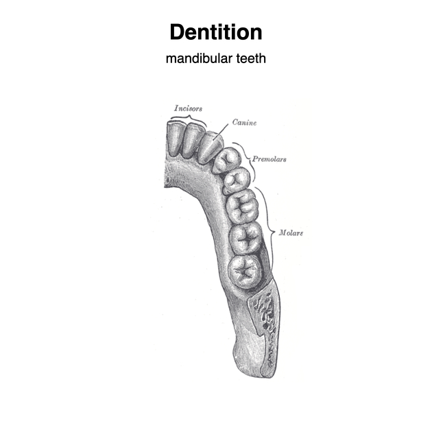
- hollowed for reception of teeth (normally 16)
- attachment of buccinator muscle
- inferior border
- rounded, longer than the superior border and thicker in front than behind
- groove for the facial artery may be present at the point it joins the ramus of the mandible
Ramus
The ramus is quadrilateral in shape, and has two surfaces, four borders, and two processes and one canal:
- external (or lateral) surface
- flat; gives attachement to the masseter muscle
- inner (or medial) surface
- mandibular foramen: inferior alveolar vessels and nerve pass into
- lingula mandible: prominent, sharp ridge in front of the mandibular foramen; gives attachment to the sphenomandibular ligament
- mandibular foramen: inferior alveolar vessels and nerve pass into
- lower border
- thick, straight and continuous with the inferior border of the body of the mandible
- posterior border
- thick, smooth, rounded and covered by the parotid gland
- angle of the mandible is at its junction of the posterior border and the body
- anterior border
- thin above and thicker below; continuous with the oblique line
- upper border
- thin
- consists of the coronoid process anteriorly and the condylar process posterior separated by the mandibular notch
The mandibular canal runs obliquely downward and forward in the ramus, and then horizontally forward in the body, where it is placed under the alveoli and communicates with them by small openings. On arriving at the incisor teeth, it turns back to communicate with the mental foramen, giving off two small canals which run to the cavities containing the incisor teeth. It contains the inferior alveolar vessels and nerve, from which branches are distributed to the teeth via the incisive nerve.
Coronoid Process
- thin, triangular eminence from the upper border of the ramus of the mandible
- separated from the condylar process posteriorly by the mandibular notch
- temporalis muscle inserts into its medial and lateral surfaces
- masseter muscle also inserts into its lateral surface
Condylar Process
- thicker than the coronoid process
- consists of two portions: condyle and neck
- articular surface for articulation with the articular disk of the temporomandibular joint (TMJ)
- convex from before backward and from side to side, and extends farther on the posterior than on the anterior surface
- condylar neck is flattened from front to back; lateral pterygoid muscle inserts into it
Blood supply
- facial artery (branch of external carotid artery)
- lingual artery (branch of external carotid artery)
- inferior alveolar artery (branch of maxillary artery)
References changed:
- 1. Susan Standring. Gray's Anatomy. (2015) ISBN: 9780702052309 - <a href="http://books.google.com/books?vid=ISBN9780702052309">Google Books</a>
- 2. H. Ric Harnsberger, André J. Macdonald. Diagnostic and Surgical Imaging Anatomy. (2006) ISBN: 9781931884297 - <a href="http://books.google.com/books?vid=ISBN9781931884297">Google Books</a>
- Gray's Anatomy 20th Edition
- Harnsberger HR, Osborn AG, Ross J et-al. Diagnostic and Surgical Imaging Anatomy. Lippincott Williams & Wilkins. (2006) ISBN:1931884293. <a href="http://books.google.com/books?vid=ISBN1931884293">Read it at Google Books</a> - <a href="http://www.amazon.com/gp/product/1931884293">Find it at Amazon</a><span class="ref_v3"></span>


 Unable to process the form. Check for errors and try again.