
Maxillary sinus
Citation, DOI, disclosures and article data
At the time the article was created Maxime St-Amant had no recorded disclosures.
View Maxime St-Amant's current disclosuresAt the time the article was last revised Abdus Sattar had no financial relationships to ineligible companies to disclose.
View Abdus Sattar's current disclosures- Maxillary antrum
- Maxillary antra
- Antrum of Highmore
- antrum Highmorianum
- Antra of Highmore
- Maxillary sinuses
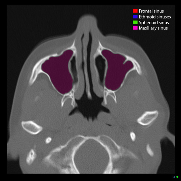
The maxillary sinus (or antrum of Highmore) is a paired pyramid-shaped paranasal sinus within the maxillary bone which drains via the maxillary ostium into the infundibulum, then through hiatus semilunaris into the middle meatus. It is the largest of the paranasal sinuses.
On this page:
Summary
location: paired sinuses within the body of the maxilla
blood supply: small arteries from the facial, maxillary, infraorbital and greater palatine arteries
innervation: anterior, middle and posterior superior alveolar, greater palatine and infraorbital nerves
Gross anatomy
Described as a pyramid, the maxillary sinuses have a base on the lateral border of the nose, with the apex pointing towards the zygomatic process of the maxilla. The floor is formed by the alveolar process of the maxilla. The roof is the orbital floor. The posterior wall forms the anterior border of the pterygopalatine fossa.
There are several recesses of the maxillary sinus 5:
infraorbital recess (superiorly)
zygomatic recess (laterally)
alveolar recess (inferiorly)
palatine recess (variable extension of alveolar recess)
Like the other paranasal air sinuses, these can vary in size. Large maxillary sinuses can extend to the alveolar process of the maxilla to the point where the roots of the molar teeth can project into the space.
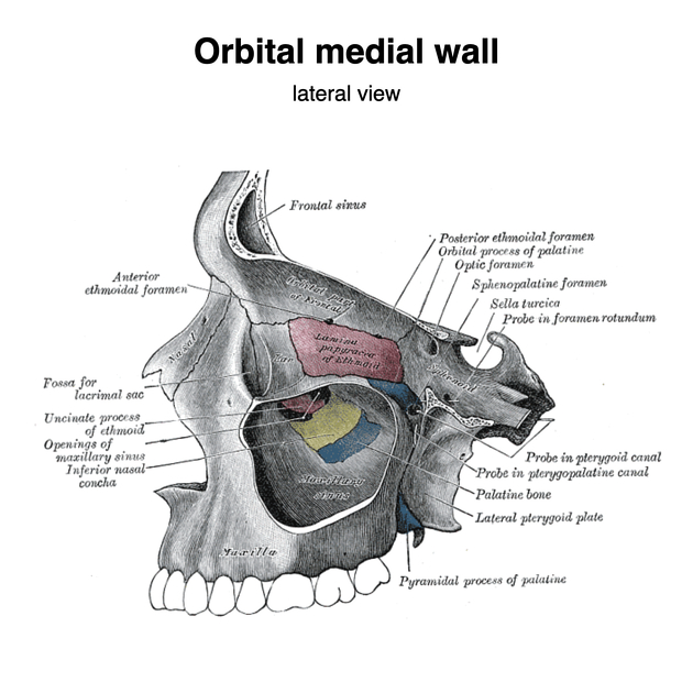
Unlike the other paranasal air sinuses, the opening of the sinus is found on its superior end. This ostium communicates with the nasal cavity via the posterior end of the hiatus semilunaris.
ADVERTISEMENT: Supporters see fewer/no ads
Arterial supply
Small arteries from the facial, maxillary, infraorbital and greater palatine arteries pierce the bony walls of the maxillary sinus.
Venous drainage
Venous drainage anteriorly is via the sphenopalatine vein and posteriorly via the pterygoid venous plexus and the facial vein.
Lymphatic drainage
Lymph from the maxillary sinus drains to the submandibular group of lymph nodes via the infraorbital foramen or the aforementioned communication with the nasal cavity.
Innervation
-
superior alveolar nerves
posterior superior alveolar nerves: dental branches pierce the bone to supply the sinus mucosa
middle superior alveolar nerves: supply the pre-molar teeth and overlying mucosa of the sinus
anterior superior alveolar nerves: supplies canine and incisors and anterior wall of the sinus
-
minute branches from this nerve supply the medial wall of the sinus
-
perforating branches supply the roof of the sinus
Variant anatomy
Common anatomic variations of maxillary sinuses are 6:
pneumatisation into the roots of teeth (83.2%)
antral septations (44.4%)
hypoplasia (4.8%)
exostosis (2.6%)
total maxillary sinus agenesis - rare
ADVERTISEMENT: Supporters see fewer/no ads
Development
It is present at birth and it develops until around the age of 14 years.
History and etymology
The antrum Highmorianum was first described by Nathaniel Highmore (1613-1685), a British physician and anatomist 3.
Related pathology
-
neoplastic
-
infections and inflammation
-
more than 70% of unilateral maxillary sinusitis can be attributed to an odontogenic cause 4
-
-
others
References
- 1. S. Jacob. Atlas of Human Anatomy. (2001) - Google Books
- 2. Mcminn. Last's Anatomy. (2003) ISBN: 9780729537520 - Google Books
- 3. Prideaux D. A Note on Nathaniel Highmore, M.D. [1613-1685], and His Memorial Tablet in Purse Caundle Church, Dorset. Proc R Soc Med. 1914;7(Sect Hist Med):106-8. PMC2003558 - Pubmed
- 4. Loureiro R, Naves E, Zanello R, Sumi D, Gomes R, Daniel M. Dental Emergencies: A Practical Guide. Radiographics. 2019;39(6):1782-95. doi:10.1148/rg.2019190019 - Pubmed
- 5. Whyte A & Boeddinghaus R. The Maxillary Sinus: Physiology, Development and Imaging Anatomy. Dentomaxillofac Radiol. 2019;48(8):20190205. doi:10.1259/dmfr.20190205 - Pubmed
- 6. Pelinsari Lana J, Moura Rodrigues Carneiro P, de Carvalho Machado V, Eduardo Alencar de Souza P, Ricardo Manzi F, Campolina Rebello Horta M. Anatomic Variations and Lesions of the Maxillary Sinus Detected in Cone Beam Computed Tomography for Dental Implants. Clin Oral Impl Res. 2011;23(12):1398-403. doi:10.1111/j.1600-0501.2011.02321.x - Pubmed
Incoming Links
- Lund-Mackay score
- Maxillary sinus mucocele
- Anterior superior alveolar nerve
- Odontogenic keratocyst
- Anterior superior alveolar artery
- Middle superior alveolar artery
- Middle meatus
- Antrolith
- Posterior superior alveolar nerve
- Maxilla
- Ethmoid bone
- Infratemporal fossa
- Pterygopalatine fossa
- Hiatus semilunaris
- Juvenile nasopharyngeal angiofibroma
- Infraorbital foramen
- Apical periodontitis
- McGrigor-Campbell lines
- Anterior superior alveolar canal
- Middle superior alveolar nerve
- Ectopic tooth in maxillary antrum with secondary chronic sinusitis
- Ruptured globe
- Antrochoanal polyp
- Pott puffy tumor
- Paranasal sinus development (Gray's illustration)
- Antrochoanal polyp
- Esthesioneuroblastoma
- Protrusion of the infraorbital canal into the maxillary sinus
- Protrusion of the infraorbital canal into the maxillary sinus
- Sinonasal nonkeratinizing squamous cell carcinoma
- Temporoparietal fracture with intracranial hemorrhage
- Sinonasal angiomatous polyp
- Recurrent dacryoadenitis
- Orbital floor blow-out fracture and ocular globe rupture
- Orbital medial wall and floor blow-out fracture
- Orbital medial wall blow-out fracture
- Adenoid cystic carcinoma of maxillary sinus
- Orbitotomy in Graves ophthalmopathy
- Chondrosarcoma of the maxilla
- Orbital medial wall blow-out fracture
Related articles: Anatomy: Head and neck
- skeleton of the head and neck
-
cranial vault
- scalp (mnemonic)
- fontanelle
-
sutures
- calvarial
- facial
- frontozygomatic suture
- frontomaxillary suture
- frontolacrimal suture
- frontonasal suture
- temporozygomatic suture
- zygomaticomaxillary suture
- parietotemporal suture (parietomastoid suture)
- occipitotemporal suture (occipitomastoid suture)
- sphenofrontal suture
- sphenozygomatic suture
- spheno-occipital suture (not a true suture)
- lacrimomaxillary suture
- nasomaxillary suture
- internasal suture
- basal/internal
- skull landmarks
- frontal bone
- temporal bone
- parietal bone
- occipital bone
- skull base (foramina)
-
facial bones
- midline single bones
- paired bilateral bones
- cervical spine
- hyoid bone
- laryngeal cartilages
-
cranial vault
- muscles of the head and neck
- muscles of the tongue (mnemonic)
- muscles of mastication
-
facial muscles
- epicranius muscle
- circumorbital and palpebral muscles
- nasal muscles
-
buccolabial muscles
- elevators, retractors and evertors of the upper lip
- levator labii superioris alaeque nasalis muscle
- levator labii superioris muscle
- zygomaticus major muscle
- zygomaticus minor muscle
- levator anguli oris muscle
- malaris muscle
- risorius muscle
- depressors, retractors and evertors of the lower lip
- depressor labii inferioris muscle
- depressor anguli oris muscle
- mentalis muscle
- compound sphincter
-
orbicularis oris muscle
- incisivus labii superioris muscle
- incisivus labii inferioris muscle
-
orbicularis oris muscle
- muscle of mastication
- modiolus
- elevators, retractors and evertors of the upper lip
- muscles of the middle ear
- orbital muscles
- muscles of the soft palate
- pharyngeal muscles
- suprahyoid muscles
- infrahyoid muscles
- intrinsic muscles of the larynx
- muscles of the neck
- platysma muscle
- longus colli muscle
- longus capitis muscle
- scalenus anterior muscle
- scalenus medius muscle
- scalenus posterior muscle
- scalenus pleuralis muscle
- sternocleidomastoid muscle
-
suboccipital muscles
- rectus capitis posterior major muscle
- rectus capitis posterior minor muscle
- obliquus capitis superior muscle
- obliquus capitis inferior muscle
- accessory muscles of the neck
- deep cervical fascia
-
deep spaces of the neck
- anterior cervical space
- buccal space
- carotid space
- danger space
- deep cervical fascia
- infratemporal fossa
- masticator space
- parapharyngeal space
- stylomandibular tunnel
- parotid space
- pharyngeal (superficial) mucosal space
- perivertebral space
- posterior cervical space
- pterygopalatine fossa
- retropharyngeal space
- suprasternal space (of Burns)
- visceral space
- surgical triangles of the neck
- orbit
- ear
- paranasal sinuses
- upper respiratory tract
- viscera of the neck
- blood supply of the head and neck
-
arterial supply
-
common carotid artery
- carotid body
- carotid bifurcation
- subclavian artery
- variants
-
common carotid artery
- venous drainage
-
arterial supply
- innervation of the head and neck
-
cranial nerves
- olfactory nerve (CN I)
- optic nerve (CN II)
- oculomotor nerve (CN III)
- trochlear nerve (CN IV)
-
trigeminal nerve (CN V) (mnemonic)
- trigeminal ganglion
- ophthalmic division
- maxillary division
- mandibular division
- abducens nerve (CN VI)
- facial nerve (CN VII)
-
vestibulocochlear nerve (CN VIII)
- vestibular ganglion (Scarpa's ganglion)
- glossopharyngeal nerve (CN IX)
- vagus nerve (CN X)
- (spinal) accessory nerve (CN XI)
- hypoglossal nerve (CN XII)
- parasympathetic ganglia of the head and neck
- cervical sympathetic ganglia
- greater occipital nerve
- third occipital nerve
-
cervical plexus
- muscular branches
- longus capitis
- longus colli
- scalenes
- geniohyoid
- thyrohyoid
-
ansa cervicalis
- omohyoid (superior and inferior bellies separately)
- sternothyroid
- sternohyoid
- phrenic nerve
- contribution to the accessory nerve (CN XI)
- cutaneous branches
- muscular branches
- brachial plexus
- pharyngeal plexus
-
cranial nerves
- lymphatic drainage of the head and neck
- embryological development of the head and neck


 Unable to process the form. Check for errors and try again.