
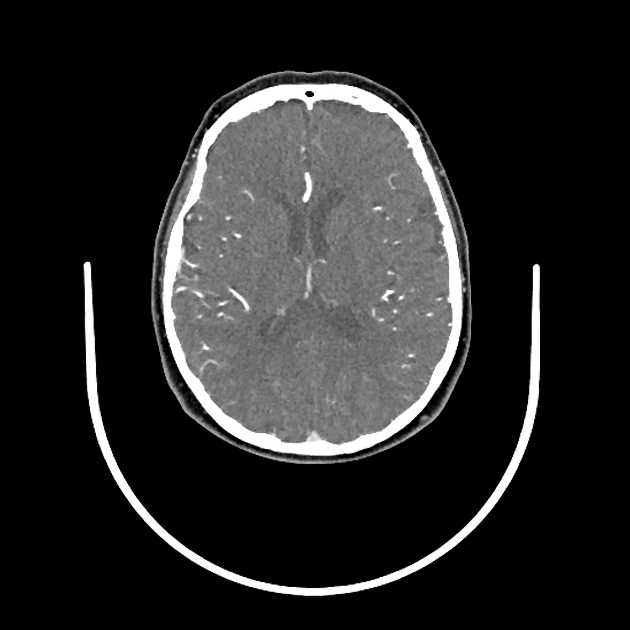
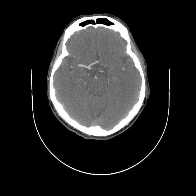
Medium vessel occlusion (MeVO), also termed distal medium vessel occlusion (DMVO) or distal vessel occlusion (DVO), describes occlusion of a medium-sized intracranial artery resulting in impending acute ischaemic stroke.
Definition
One consensus definition of ‘medium vessel’ suggests intracranial arteries with a luminal diameter of 0.75-2.0 mm 1. Thus, arteries included in the definition of medium vessel occlusion include 1-3:
M3 and M4 segments of the middle cerebral arteries
A2, A3, A4, and A5 segments of the anterior cerebral arteries
P2, P3, P4, and P5 segments of the posterior cerebral arteries
The M2 segments of the middle cerebral arteries, A1 segments of the anterior cerebral arteries and the P1 segment of the posterior cerebral arteries have heterogenous angioarchitecture among patients and as a result, may be variably defined as either sites of medium vessel occlusion or large vessel occlusion (LVO) 1-3. Please see the article on large vessel occlusion (LVO) for further discussion.
Pragmatically, randomised clinical trials of endovascular clot retrieval in medium vessel occlusion have employed the following definitions of 'medium vessel':
-
ESCAPE-MeVO trial 10:
M2 or M3 segments of the middle cerebral arteries
A2 or A3 segments of the anterior cerebral arteries
P2 or P3 segments of the posterior cerebral arteries
-
DISTAL trial 11:
nondominant or codominant M2 segment of the middle cerebral arteries
M3 or M4 segments of the middle cerebral arteries
A1, A2, or A3 segments of the anterior cerebral arteries
P1, P2, or P3 segments of the posterior cerebral arteries
Clinical importance
medium vessel occlusion accounts for 25-40% of acute ischaemic stroke 8
multi-phase CT angiography with CT perfusion is more sensitive than single-phase CT angiography to detect medium vessel occlusions 6
in CT perfusion, the traditional Tmax delay cut-off of >6 seconds may not detect perfusion defects due to medium vessel occlusion in 10% of cases, and in those cases milder Tmax delay (e.g. >4 seconds) may be present 9
although initial research was somewhat equivocal 4,5,7,8, randomised control trials have not found endovascular clot retrieval to be superior to best medical therapy in patients with acute ischaemic stroke due to medium vessel occlusion 10,11


 Unable to process the form. Check for errors and try again.