
MIBG
Citation, DOI, disclosures and article data
At the time the article was created Jeremy Jones had no recorded disclosures.
View Jeremy Jones's current disclosuresAt the time the article was last revised Arlene Campos had no financial relationships to ineligible companies to disclose.
View Arlene Campos's current disclosures- Metaiodobenzylguanidine imaging
- Metaiodobenzylguanidine scan
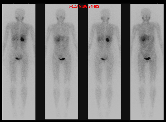
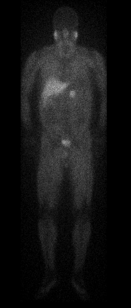
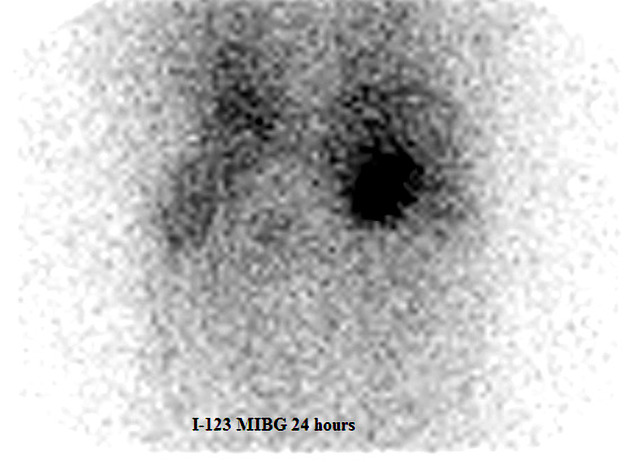
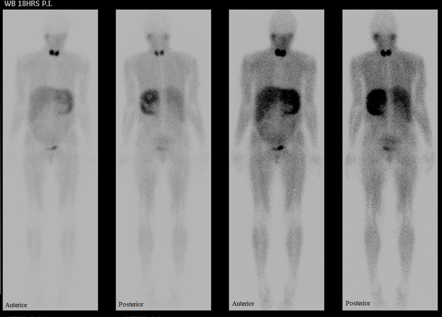
MIBG scan is a scintigraphic study that uses metaiodobenzylguanidine, noradrenaline analogue 9, labelled to iodine-123 or iodine-131. It is indicated in the investigation of phaeochromocytoma. I-131 MIBG, also called I-131 iobenguane, is a theranostic agent used to treat unresectable MIBG-positive tumours.
Indications
MIBG is positive in:
Some mild uptake can be physiologic in 4:
liver
salivary glands
urinary bladder
heart
gastrointestinal tract
adrenal glands
brown fat
Cardiac imaging studies with MIBG labelled to radio-iodine may be useful in the evaluation of cardiac toxicity from chemotherapy 5-8.
Procedure
Iodine-123 has 13 hours of half-life and allows imaging up to 48 hours. Iodine-131 is only used when iodine-123 is not available. The higher energy photons emitted by iodine-131 render it inferior to iodine-123 because of higher dose it gives to patients 9.
Medications that can interfere with MIBG uptake should be stopped such as tricyclic antidepressants, antihypertensives, and decongestant drugs such as pseudoephedrine, phenylpropanolamine, and phenylephrine 9.
To reduce radiation dose to the thyroid gland, 400 mg of oral potassium perchlorate is given at one hour before MIBG injection in adults. In children, 0.1 to 0.2 ml of Lugol's iodine diluted with water or milk is given three times a day starting at 48 hours before the injection 9.
Technique
administer intravenous MIBG slowly over 1 to 2 minutes to prevent adrenergic side effects in case of fast injection 9
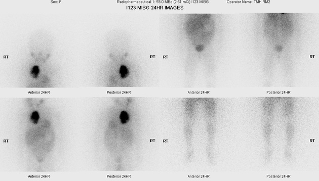
images are taken for anterior and posterior abdomen at 10 to 20 minutes per view 9
whole body image is taken to look for metastasis 9
SPECT is helpful for anatomical localisation 9
pelvic images taken at 24 hours after urinary bladder emptying. Sometimes, images may also be taken at 4 or 48 hours 9
Quiz questions
References
- 1. Sasajima T, Kinouchi H, Tomura N, Watarai J, Mizoi K. High Uptake of 123I-Metaiodobenzylguanidine Related to Olfactory Neuroblastoma Revealed by Single-Photon Emission CT. AJNR Am J Neuroradiol. 2000;21(4):717-20. PMC7976631 - Pubmed
- 2. Paltiel HJ, Gelfand MJ, Elgazzar AH et-al. Neural crest tumors: I-123 MIBG imaging in children. Radiology. 1994;190 (1): 117-21. Radiology (abstract) - Pubmed citation
- 3. Van gils AP, Falke TH, Van erkel AR et-al. MR imaging and MIBG scintigraphy of pheochromocytomas and extraadrenal functioning paragangliomas. Radiographics. 1991;11 (1): 37-57. Radiographics (abstract) - Pubmed citation
- 4. Hanson M, Feldman J, Blinder R, Moore J, Coleman R. Carcinoid Tumors: Iodine-131 MIBG Scintigraphy. Radiology. 1989;172(3):699-703. doi:10.1148/radiology.172.3.2772175 - Pubmed
- 5. Casáns-Tormo I, Jiménez-Heffernan A, Pubul-Núñez V, Ruano-Pérez R. Cardiac sympathetic innervation scintigraphy with I-meta-iodobenzylguanidine. Basis, protocols and clinical applications in Cardiology. (2019) Revista espanola de medicina nuclear e imagen molecular. 38 (4): 262-271. doi:10.1016/j.remn.2019.01.001 - Pubmed
- 6. Laursen A, Ripa R, Hasbak P et al. J Nucl Cardiol. 2020;27(3):931-9. doi:10.1007/s12350-018-01566-y - Pubmed
- 7. Laursen AH, Thune JJ, Hutchings M, Hasbak P, Kjaer A, Elming MB, Ripa RS. I-MIBG imaging for detection of anthracycline-induced cardiomyopathy. (2018) Clinical physiology and functional imaging. 38 (2): 176-185. doi:10.1111/cpf.12419 - Pubmed
- 8. Dos Santos M, da Rocha E, Verberne H, da Silva E, Aragon D, Junior J. Assessment of Late Anthracycline-Induced Cardiotoxicity by I-MIBG Cardiac Scintigraphy in Patients Treated During Childhood and Adolescence. J Nucl Cardiol. 2017;24(1):256-64. doi:10.1007/s12350-015-0309-y - Pubmed
- 9. Guide to Radiological Procedures. (2009) - Google Books
Incoming Links
Related articles: Imaging technology
- imaging technology
- imaging physics
- imaging in practice
-
x-rays
- x-ray physics
- x-ray in practice
- x-ray production
- x-ray tube
- filters
- automatic exposure control (AEC)
- beam collimators
- grids
- air gap technique
- cassette
- intensifying screen
- x-ray film
- image intensifier
- digital radiography
- digital image
- mammography
- x-ray artifacts
- radiation units
- radiation safety
- radiation detectors
- fluoroscopy
-
computed tomography (CT)
- CT physics
- CT in practice
- CT technology
- CT image reconstruction
- CT image quality
- CT dose
-
CT contrast media
-
iodinated contrast media
- agents
- water soluble
- water insoluble
- vicarious contrast material excretion
- iodinated contrast media adverse reactions
- agents
- non-iodinated contrast media
-
iodinated contrast media
-
CT artifacts
- patient-based artifacts
- physics-based artifacts
- hardware-based artifacts
- ring artifact
- tube arcing
- out of field artifact
- air bubble artifact
- helical and multichannel artifacts
- CT safety
- history of CT
-
MRI
- MRI physics
- MRI in practice
- MRI hardware
- signal processing
-
MRI pulse sequences (basics | abbreviations | parameters)
- T1 weighted image
- T2 weighted image
- proton density weighted image
- chemical exchange saturation transfer
- CSF flow studies
- diffusion weighted imaging (DWI)
- echo-planar pulse sequences
- fat-suppressed imaging sequences
- gradient echo sequences
- inversion recovery sequences
- metal artifact reduction sequence (MARS)
-
perfusion-weighted imaging
- techniques
- derived values
- saturation recovery sequences
- spin echo sequences
- spiral pulse sequences
- susceptibility-weighted imaging (SWI)
- T1 rho
- MR angiography (and venography)
-
MR spectroscopy (MRS)
- 2-hydroxyglutarate peak: resonates at 2.25 ppm
- alanine peak: resonates at 1.48 ppm
- choline peak: resonates at 3.2 ppm
- citrate peak: resonates at 2.6 ppm
- creatine peak: resonates at 3.0 ppm
- functional MRI (fMRI)
- gamma-aminobutyric acid (GABA) peak: resonates at 2.2-2.4 ppm
- glutamine-glutamate peak: resonates at 2.2-2.4 ppm
- Hunter's angle
- lactate peak: resonates at 1.3 ppm
- lipids peak: resonates at 1.3 ppm
- myoinositol peak: resonates at 3.5 ppm
- MR fingerprinting
- N-acetylaspartate (NAA) peak: resonates at 2.0 ppm
- propylene glycol peak: resonates at 1.13 ppm
-
MRI artifacts
- MRI hardware and room shielding
- MRI software
- patient and physiologic motion
- tissue heterogeneity and foreign bodies
- Fourier transform and Nyquist sampling theorem
- MRI contrast agents
- MRI safety
-
ultrasound
- ultrasound physics
-
transducers
- linear array
- convex array
- phased array
- frame averaging (frame persistence)
- ultrasound image resolution
- imaging modes and display
- pulse-echo imaging
- real-time imaging
-
Doppler imaging
- Doppler effect
- colour Doppler
- power Doppler
- B flow
- colour box
- Doppler angle
- pulse repetition frequency and scale
- wall filter
- colour write priority
- packet size (dwell time)
- peak systolic velocity
- end-diastolic velocity
- resistive index
- pulsatility index
- Reynolds number
- panoramic imaging
- compound imaging
- harmonic imaging
- elastography
- scanning modes
- 2D ultrasound
- 3D ultrasound
- 4D ultrasound
- M-mode
-
ultrasound artifacts
- acoustic shadowing
- acoustic enhancement
- beam width artifact
- reverberation artifact
- ring down artifact
- mirror image artifact
- side lobe artifact
- speckle artifact
- speed displacement artifact
- refraction artifact
- multipath artifact
- anisotropy
- electrical interference artifact
- hardware-related artifacts
- Doppler artifacts
- aliasing
- tissue vibration
- spectral broadening
- blooming
- motion (flash) artifact
- twinkling artifact
- acoustic streaming
- biological effects of ultrasound
- history of ultrasound
-
nuclear medicine
- nuclear medicine physics
- detectors
- tissue to background ratio
-
radiopharmaceuticals
- fundamentals of radiopharmaceuticals
- radiopharmaceutical labelling
- radiopharmaceutical production
- nuclear reactor produced radionuclides
- cyclotron produced radionuclides
- radiation detection
- dosimetry
- specific agents
- carbon-11
- chromium-51
- fluorine agents
- gallium agents
- Ga-67 citrate
- Ga-68
- iodine agents
-
I-123
- I-123 iodide
- I-123 ioflupane (DaTSCAN)
- I-123 ortho-iodohippurate
- I-131
-
MIBG scans
- I-123 MIBG
- I-131 MIBG
-
I-123
- indium agents
- In-111 Octreoscan
- In-111 OncoScint
- In-111 Prostascint
- In-111 oxine labelled WBC
- krypton-81m
- nitrogen-13
- oxygen-15
- phosphorus-32
- selenium-75
-
technetium agents
- Tc-99m DMSA
- Tc-99m DTPA
- Tc-99m DTPA aerosol
- Tc-99m HMPAO
- Tc-99m HMPAO labelled WBC
- Tc-99m MAA
- Tc-99m MAG3
- Tc-99m MDP
- Tc-99m mercaptoacetyltriglycine
- Tc-99m pertechnetate
- Tc-99m labelled RBC
- Tc-99m sestamibi
- Tc-99m sulfur colloid
- Tc-99m sulfur colloid (oral)
- thallium-201 chloride
- xenon agents
- in vivo therapeutic agents
- pharmaceuticals used in nuclear medicine
-
emerging methods in medical imaging
- radiography
- phase-contrast imaging
- CT
- deep-learning reconstruction
- photon counting CT
- virtual non-contrast imaging
- ultrasound
- magnetomotive ultrasound (MMUS)
- superb microvascular imaging
- ultrafast Doppler imaging
- ultrasound localisation microscopy
- MRI
- nuclear medicine
- total body PET system
- immuno-PET
- miscellaneous
- radiography


 Unable to process the form. Check for errors and try again.