
Midventricular hypertrophic cardiomyopathy is a phenotype or morphological variant of asymmetric hypertrophic cardiomyopathy (HCM) characterized by hypertrophy in the midventricular segment that might result in midventricular obstruction.
On this page:
Epidemiology
Midventricular hypertrophic cardiomyopathy is rare 1 accounting for up to 4-10% 2-4.
Association
Midventricular hypertrophic cardiomyopathy in particular with midventricular obstruction is associated with left ventricular apical aneurysms.
Clinical presentation
Symptoms of midventricular hypertrophic cardiomyopathy and midventricular obstruction do not differ significantly from other types of hypertrophic cardiomyopathy or left ventricular outflow obstruction and include performance deficits, dyspnea, angina, presyncope, syncope on exertion 2.
On auscultation, there might be an apical systolic murmur like or variably a long mitral diastolic murmur 2.
Complications
Complications of midventricular hypertrophic cardiomyopathy include 1-4:
systemic embolism
ventricular arrhythmias
Pathology
Midventricular hypertrophic cardiomyopathy is characterized by hypertrophy of the midventricular segments not only of the septum but also the free wall, which can result in midventricular obstruction.
Midventricular obstruction is defined by a peak gradient of ≥30 mmHg of the left ventricular mid cavity 1. There are apparently significant variations in the size of the obstructed apical cavity 2.
This phenotype is typically not associated with obstruction of the left ventricular outflow tract or systolic anterior motion (SAM) of the anterior leaflet of the mitral valve 1.
Radiographic features
The typical imaging feature of midventricular hypertrophic cardiomyopathy is a dumbell shape or hourglass appearance of the left ventricle, which can be conveniently appreciated on left ventricular long-axis views 1.
Ultrasound
Echocardiography
Echocardiography can demonstrate the hypertrophied left ventricular segments and can aid in the evaluation of midventricular obstruction defined by a peak mid cavity gradient ≥30 mmHg 1,4-6.
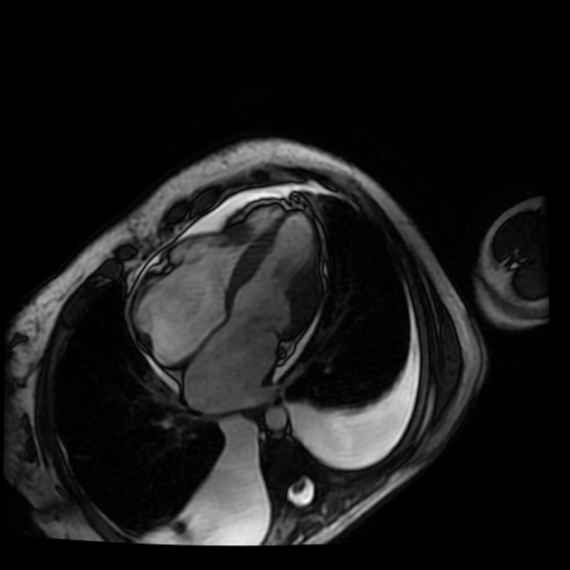
MRI
Cardiac MRI can show hypertrophy of the midventricular segments and can aid in the evaluation of cardiac volumes and cardiac function and associated myocardial fibrosis and scarring 1. The following features might be present in midventricular hypertrophic cardiomyopathy 1,3-7:
-
confined midventricular wall thickening
dumbell or hourglass shape of the left ventricle
left ventricular apical aneurysm
preserved or increased ejection fraction
-
cardiac tissue characterization
T1 mapping: increased native T1 values
ECV: increased
-
IRGE/PSIR:
late gadolinium enhancement in midventricular and apical segments
indicating replacement fibrosis or myocardial scarring
might show complications as apical aneurysms or intracardiac thrombi
Radiology report
The radiological report should include a description of the following 1-6:
location and extent of hypertrophic wall segments
cardiac volumes and measurements including left ventricular mass
signs of myocardial fibrosis, myocardial scarring or replacement fibrosis
complications as apical aneurysms or left ventricular thrombus formation
mid cavity gradient
Differential diagnosis
Conditions that might mimic the clinical presentation or imaging appearance of asymmetric hypertrophic cardiomyopathy include 1,4-6:


 Unable to process the form. Check for errors and try again.