Citation, DOI, disclosures and article data
Citation:
Sharma R, Motor band sign. Reference article, Radiopaedia.org (Accessed on 30 Mar 2025) https://doi.org/10.53347/rID-58837
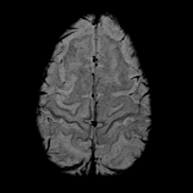
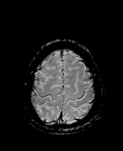
The motor band sign is a radiological sign described in amyotrophic lateral sclerosis (ALS).
It refers to the appearance of the primary motor cortex on axial GRE or SWI MRI sequences in patients with amyotrophic lateral sclerosis and some of its clinical variants 1,2,6. On these sequences, and in the axial plane, curvilinear bands of low signal may be appreciated within the cortical gray matter of the primary motor cortex (i.e. 'motor bands' of low signal) 1,2,6. Occasionally, T2/FLAIR sequences may also demonstrate the same low signal 1-4.
While this signal abnormality is usually seen along the whole primary motor cortex, in the progressive bulbar palsy variant, the signal change may only be seen laterally (i.e. in the faciobulbar region; see homunculus) 6. Furthermore, although this pattern of signal change is usually seen bilaterally, it has been reported to also occur unilaterally 2.
This sign is produced because of iron accumulation within microglia in the motor cortex 1. The mechanisms for this have not been fully elucidated, however it has been postulated that perhaps microglia accumulate this iron as they phagocytose degenerating neurones from the primary motor cortex 1. The neurones most affected are those in the middle layer of the primary motor cortex 6.
It is important to note that this radiological sign is not completely specific for amyotrophic lateral sclerosis 1-4. Indeed, low T2/FLAIR/GRE/SWI signal in the precentral gyrus has been documented to also occur in patients with other neurological disorders (e.g. Parkinson disease, Alzheimer disease, spinocerebellar ataxia type 17) and even in many normal adults as an age-related phenomenon 1-5. This might be due to the fact that the primary motor cortex has the highest cortical concentration of iron 1.
-
1. Kwan JY, Jeong SY, Van Gelderen P, Deng HX, Quezado MM, Danielian LE, Butman JA, Chen L, Bayat E, Russell J, Siddique T, Duyn JH, Rouault TA, Floeter MK. Iron accumulation in deep cortical layers accounts for MRI signal abnormalities in ALS: correlating 7 tesla MRI and pathology. (2012) PloS one. 7 (4): e35241. doi:10.1371/journal.pone.0035241 - Pubmed
-
2. Chakraborty S, Gupta A, Nguyen T, Bourque P. The "Motor Band Sign:" Susceptibility-Weighted Imaging in Amyotrophic Lateral Sclerosis. (2015) The Canadian journal of neurological sciences. Le journal canadien des sciences neurologiques. 42 (4): 260-3. doi:10.1017/cjn.2015.40 - Pubmed
-
3. Ngai S, Tang YM, Du L, Stuckey S. Hyperintensity of the precentral gyral subcortical white matter and hypointensity of the precentral gyrus on fluid-attenuated inversion recovery: variation with age and implications for the diagnosis of amyotrophic lateral sclerosis. (2007) AJNR. American journal of neuroradiology. 28 (2): 250-4. Pubmed
-
4. Imon Y, Yamaguchi S, Yamamura Y, Tsuji S, Kajima T, Ito K, Nakamura S. Low intensity areas observed on T2-weighted magnetic resonance imaging of the cerebral cortex in various neurological diseases. (1995) Journal of the neurological sciences. 134 Suppl: 27-32. Pubmed
-
5. Dash D & Mestre T. Motor Band Sign in a Huntington Disease Phenocopy. Parkinsonism & Related Disorders. 2023;:105333. doi:10.1016/j.parkreldis.2023.105333
-
6. Desai A, Agarwal A, Mohamed A et al. Motor Neuron Diseases and Central Nervous System Tractopathies: Clinical-Radiologic Correlation and Diagnostic Approach. Radiographics. 2025;45(1):e240067. doi:10.1148/rg.240067 - Pubmed
Promoted articles (advertising)


 Unable to process the form. Check for errors and try again.