
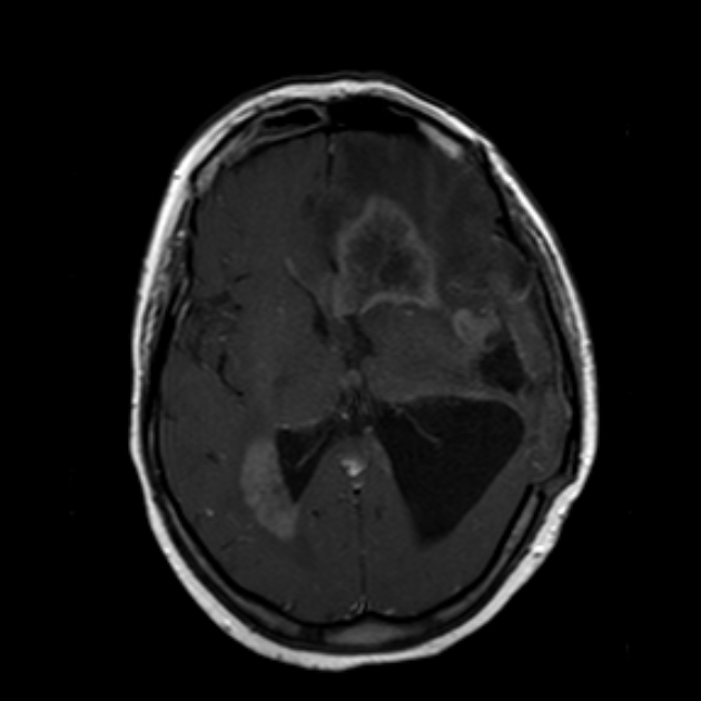
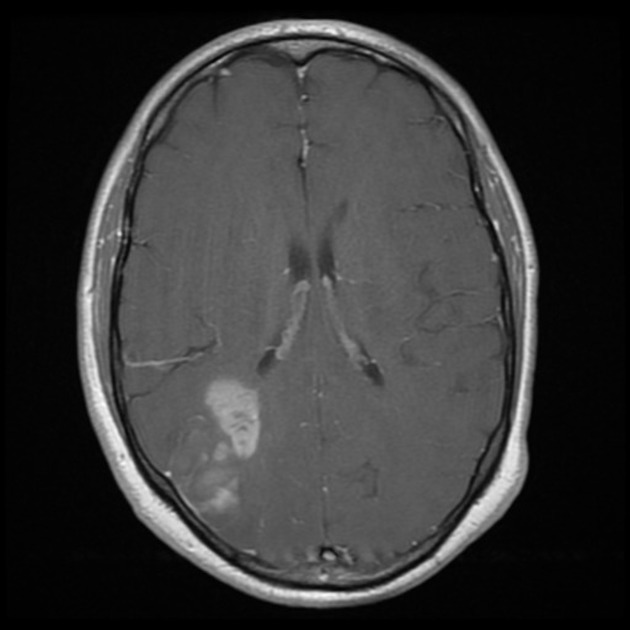
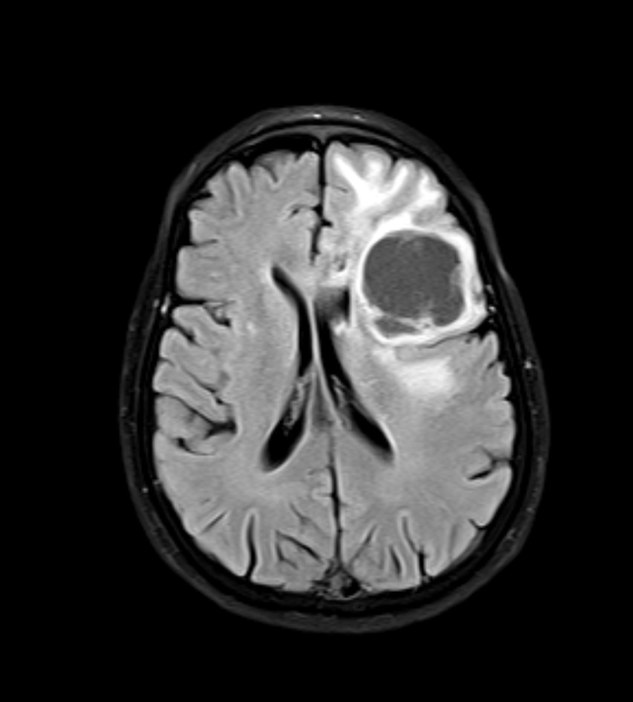
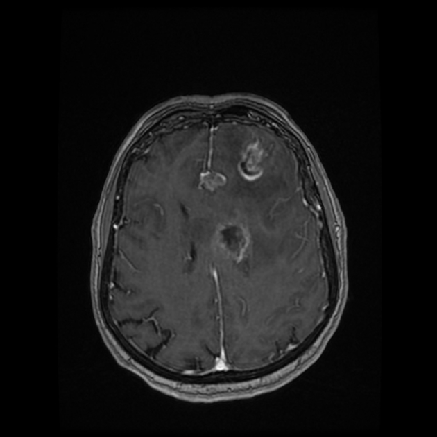
Multifocal glioblastoma
Citation, DOI, disclosures and article data
At the time the article was created Frank Gaillard had no recorded disclosures.
View Frank Gaillard's current disclosuresAt the time the article was last revised Bálint Botz had no recorded disclosures.
View Bálint Botz's current disclosures- Multifocal glioblastomas
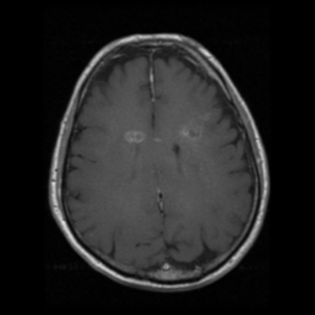
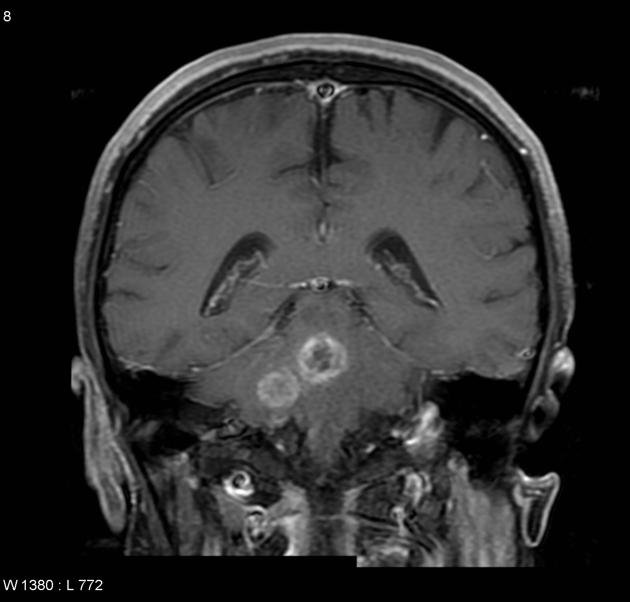
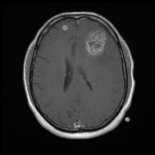
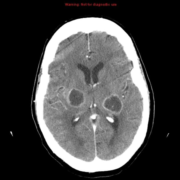
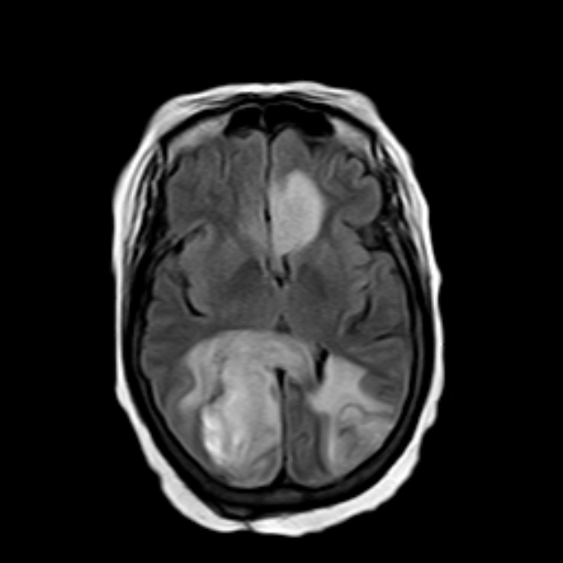
Multifocal glioblastomas are tumors which have multiple discrete areas of contrast-enhancing tumor embedded with, or connected by, T2/FLAIR signal abnormality. Multifocal glioblastomas are considered to be part of the one tumor and are commonly encountered, accounting for 2-20% of all glioblastomas 3,4.
Multifocal glioblastomas have been shown to have a poorer prognosis than solitary tumors 2,3.
Differential diagnosis
This is in contrast to multicentric glioblastomas, which have enhancing foci with normal intervening brain, and are thought to more likely represent synchronous but separate tumors.
References
- 1. Zamponi N, Rychlicki F, Ducati A et-al. Multicentric glioma with unusual clinical presentation. Childs Nerv Syst. 2001;17 (1-2): 101-5. Pubmed citation
- 2. Hassaneen W, Levine NB, Suki D et-al. Multiple craniotomies in the management of multifocal and multicentric glioblastoma. Clinical article. J. Neurosurg. 2011;114 (3): 576-84. doi:10.3171/2010.6.JNS091326 - Pubmed citation
- 3. Patil CG, Yi A, Elramsisy A, Hu J, Mukherjee D, Irvin DK, Yu JS, Bannykh SI, Black KL, Nuño M. Prognosis of patients with multifocal glioblastoma: a case-control study. (2012) Journal of neurosurgery. 117 (4): 705-11. doi:10.3171/2012.7.JNS12147 - Pubmed
- 4. Singh G, Mehrotra A, Sardhara J, Das KK, Jamdar J, Pal L, Srivastava AK, Sahu RN, Jaiswal AK, Behari S. Multiple glioblastomas: Are they different from their solitary counterparts?. (2015) Asian journal of neurosurgery. 10 (4): 266-71. doi:10.4103/1793-5482.162685 - Pubmed
Incoming Links
Related articles: Astrocytic tumour
-
astrocytic tumors
- WHO classification of CNS tumors
- WHO grading of CNS tumors
- VASARI MRI feature set
- diffuse astrocytoma grading[+][+]
- grade I: [+][+]
- grade II: [+][+]
- chordoid glioma of the third ventricle
-
low-grade diffuse astrocytoma
- fibrillary astrocytoma (no longer recognized)
- protoplasmic astrocytoma (no longer recognized)
- gemistocytic astrocytoma
- oligoastrocytoma (no longer recognized)
- pilomyxoid astrocytoma
- pleomorphic xanthoastrocytoma
- grade III[+][+]
- anaplastic astrocytoma (no longer recognized)
- anaplastic oligoastrocytoma (no longer recognized)
- anaplastic pleomorphic xanthoastrocytoma (no longer recognized)
- grade IV: [+][+]
-
glioblastoma (GBM)
- glioblastoma IDH wildtype
- glioblastoma IDH mutant
- glioblastoma NOS
- variants
- diffuse midline glioma H3 K27M–mutant
-
glioblastoma (GBM)
- glioblastoma vs cerebral metastasis
- radiation-induced gliomas
- gliomatosis cerebri (growth pattern)
- specific locations[+][+]
- treatment response
- prognostic genetic markers[+][+]


 Unable to process the form. Check for errors and try again.