
Nasolacrimal drainage apparatus
Citation, DOI, disclosures and article data
At the time the article was created Henry Knipe had no recorded disclosures.
View Henry Knipe's current disclosuresAt the time the article was last revised Kajanan Nithiyananthan had no financial relationships to ineligible companies to disclose.
View Kajanan Nithiyananthan's current disclosures- Nasolacrimal apparatus
The nasolacrimal (drainage) apparatus consists of:
Gross anatomy
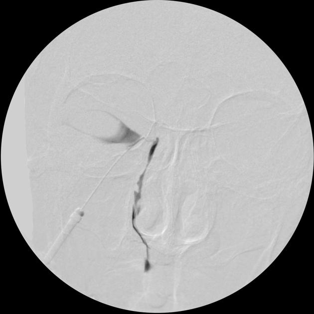
Tears produced by the lacrimal gland, accessory lacrimal glands of Krause, Wolfring and Zeis, and Meibomian glands track medially along the eyelid margins and collect at the lacrimal lake at the inner canthus. The nasolacrimal drainage apparatus drains tears in a descending course through the inferior and superior lacrimal canaliculi and common canaliculus through the valve of Rosenmuller leading to the lacrimal sac, which drains into the nasolacrimal duct within the bony nasolacrimal canal of the maxilla and through the valve of Hasner to the inferior nasal meatus below the inferior turbinate in the nasal cavity.
Note that the nasolacrimal apparatus is not to be confused with the lacrimal apparatus, which also includes the lacrimal gland.
Related pathology
-
squamous cell papillomas and carcinomas
transitional cell papillomas and carcinomas
oncocytomas
adenocarcinoma
adenoid cystic carcinoma
malignant melanomas
References
- 1. Russell EJ, Czervionke L, Huckman M et-al. CT of the inferomedial orbit and the lacrimal drainage apparatus: normal and pathologic anatomy. AJR Am J Roentgenol. 1985;145 (6): 1147-54. doi:10.2214/ajr.145.6.1147 - Pubmed citation
- 2. Jinkins JR. Atlas of Neuroradiologic Embryology, Anatomy, and Variants. Lippincott Williams & Wilkins. (2000) ISBN:0781716527. Read it at Google Books - Find it at Amazon
- 3. Kumar VA, Esmaeli B, Ahmed S et-al. Imaging Features of Malignant Lacrimal Sac and Nasolacrimal Duct Tumors. AJNR Am J Neuroradiol. doi:10.3174/ajnr.A4882 - Pubmed citation
- 4. Osama A. Raslan, Arzu Ozturk, Nancy Pham, Jennifer Chang, Edward B. Strong, Matthew Bobinski. A Comprehensive Review of Cross-Sectional Imaging of the Nasolacrimal Drainage Apparatus: What Radiologists Need to Know. (2019) American Journal of Roentgenology. doi:10.2214/AJR.19.21507 - Pubmed
Incoming Links
- Nasolacrimal tube complicated by infection
- Dacryocystitis
- Dacryocystocele
- Dacryocystocele
- Dacryocystitis and dacryocystocele
- Dacryocystocele
- Nasolacrimal duct obstruction
- Lester Jones tube
- Bilateral nasolacrimal duct mucocele and dacryocystocele
- Dacryocystocele
- Post-saccular lacrimal duct stenosis
- Dacryocystocele - acquired
- Dacryocystocoele
Related articles: Anatomy: Head and neck
- skeleton of the head and neck
-
cranial vault
- scalp (mnemonic)
- fontanelle
-
sutures
- calvarial
- facial
- frontozygomatic suture
- frontomaxillary suture
- frontolacrimal suture
- frontonasal suture
- temporozygomatic suture
- zygomaticomaxillary suture
- parietotemporal suture (parietomastoid suture)
- occipitotemporal suture (occipitomastoid suture)
- sphenofrontal suture
- sphenozygomatic suture
- spheno-occipital suture (not a true suture)
- lacrimomaxillary suture
- nasomaxillary suture
- internasal suture
- basal/internal
- skull landmarks
- frontal bone
- temporal bone
- parietal bone
- occipital bone
- skull base (foramina)
-
facial bones
- midline single bones
- paired bilateral bones
- cervical spine
- hyoid bone
- laryngeal cartilages
-
cranial vault
- muscles of the head and neck
- muscles of the tongue (mnemonic)
- muscles of mastication
-
facial muscles
- epicranius muscle
- circumorbital and palpebral muscles
- nasal muscles
-
buccolabial muscles
- elevators, retractors and evertors of the upper lip
- levator labii superioris alaeque nasalis muscle
- levator labii superioris muscle
- zygomaticus major muscle
- zygomaticus minor muscle
- levator anguli oris muscle
- malaris muscle
- risorius muscle
- depressors, retractors and evertors of the lower lip
- depressor labii inferioris muscle
- depressor anguli oris muscle
- mentalis muscle
- compound sphincter
-
orbicularis oris muscle
- incisivus labii superioris muscle
- incisivus labii inferioris muscle
-
orbicularis oris muscle
- muscle of mastication
- modiolus
- elevators, retractors and evertors of the upper lip
- muscles of the middle ear
- orbital muscles
- muscles of the soft palate
- pharyngeal muscles
- suprahyoid muscles
- infrahyoid muscles
- intrinsic muscles of the larynx
- muscles of the neck
- platysma muscle
- longus colli muscle
- longus capitis muscle
- scalenus anterior muscle
- scalenus medius muscle
- scalenus posterior muscle
- scalenus pleuralis muscle
- sternocleidomastoid muscle
-
suboccipital muscles
- rectus capitis posterior major muscle
- rectus capitis posterior minor muscle
- obliquus capitis superior muscle
- obliquus capitis inferior muscle
- accessory muscles of the neck
- deep cervical fascia
-
deep spaces of the neck
- anterior cervical space
- buccal space
- carotid space
- danger space
- deep cervical fascia
- infratemporal fossa
- masticator space
- parapharyngeal space
- stylomandibular tunnel
- parotid space
- pharyngeal (superficial) mucosal space
- perivertebral space
- posterior cervical space
- pterygopalatine fossa
- retropharyngeal space
- suprasternal space (of Burns)
- visceral space
- surgical triangles of the neck
- orbit
- ear
- paranasal sinuses
- upper respiratory tract
- viscera of the neck
- blood supply of the head and neck
-
arterial supply
-
common carotid artery
- carotid body
- carotid bifurcation
- subclavian artery
- variants
-
common carotid artery
- venous drainage
-
arterial supply
- innervation of the head and neck
-
cranial nerves
- olfactory nerve (CN I)
- optic nerve (CN II)
- oculomotor nerve (CN III)
- trochlear nerve (CN IV)
-
trigeminal nerve (CN V) (mnemonic)
- trigeminal ganglion
- ophthalmic division
- maxillary division
- mandibular division
- abducens nerve (CN VI)
- facial nerve (CN VII)
-
vestibulocochlear nerve (CN VIII)
- vestibular ganglion (Scarpa's ganglion)
- glossopharyngeal nerve (CN IX)
- vagus nerve (CN X)
- (spinal) accessory nerve (CN XI)
- hypoglossal nerve (CN XII)
- parasympathetic ganglia of the head and neck
- cervical sympathetic ganglia
- greater occipital nerve
- third occipital nerve
-
cervical plexus
- muscular branches
- longus capitis
- longus colli
- scalenes
- geniohyoid
- thyrohyoid
-
ansa cervicalis
- omohyoid (superior and inferior bellies separately)
- sternothyroid
- sternohyoid
- phrenic nerve
- contribution to the accessory nerve (CN XI)
- cutaneous branches
- muscular branches
- brachial plexus
- pharyngeal plexus
-
cranial nerves
- lymphatic drainage of the head and neck
- embryological development of the head and neck


 Unable to process the form. Check for errors and try again.