
Nasolacrimal duct
Citation, DOI, disclosures and article data
At the time the article was created Henry Knipe had no recorded disclosures.
View Henry Knipe's current disclosuresAt the time the article was last revised Arlene Campos had no financial relationships to ineligible companies to disclose.
View Arlene Campos's current disclosures- Naso-lacrimal duct
- Valve of Krause
- Nasolacrimal duct (NLD)
- Valve of Hasner
- Valve of Taillefer
- Valve of Béraud
- Valve of Huschke
- Valve of Rosenmüller
- Valve of Bochdalek
- Valve of Foltz
- Valve of Cruveilhier
- Valve of Bianchi
The nasolacrimal duct (NLD) is the terminal part of the nasolacrimal apparatus.
Gross anatomy
The nasolacrimal duct is the inferior continuation of the lacrimal sac and is ~17 mm in length in total. The duct runs in the bony nasolacrimal canal. There are two parts to the nasolacrimal duct:
intraosseous part (12 mm): enters the lacrimal groove and descends within the nasolacrimal canal of the maxilla
membranous part (5 mm): runs in the nasal mucosa; terminates below the inferior nasal meatus as a slit-like opening where it is covered by a mucosal fold called the valve of Hasner (or plica lacrimalis)
Valves
Up to eight 'valves' of the nasolacrimal duct have been defined in the literature and are commonly seen in older anatomical texts and monographs. However, it has been contended that some of these are not true mucosal valves but just embryological irregularities in the ductal wall. From craniad to caudad, these 'valves' have been named: Foltz, Bochdalek, Rosenmüller, Huschke, Aubaret, Krause (or Béraud), Taillefer, Hasner (or Cruveilhier/Bianchi) 3,4
The valves of Bochdalek and Foltz are located within the lacrimal puncta, the valves of Rosenmuller and Huschke control the ostium of canaliculi to the lacrimal sac, the valve of Krause guards the path where the lacrimal sac enters the nasolacrimal duct, and the valve of Teileffer is located in the central lacrimal duct 5.
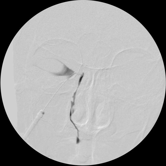
Therefore a retrospective study used digital subtraction dacryocystography (a high spatial resolution technique) to precisely define the intraluminal anatomy of the nasolacrimal duct and determine which of these named structures really represent consistent anatomical features. The researchers used a study group of 92 individuals for whom DS-DCG had previously been reported as normal 3,4:
The inferior group of 'valves' were consistently better seen:
valve of Krause: was visible in 79.3% cases
valve of Taillefer: 93.5% cases
valve of Hasner (plica lacrimalis): 98.9% cases
The superior group of 'valves' were inconsistently visualized and were therefore thought to be mucosal irregularities rather than true anatomical valves:
valve of Foltz and valve of Bochdalek: were visible in 17.1% cases
valve of Rosenmüller and valve of Huschke: 46.4% cases
valve of Aubaret: 40% cases
Related pathology
References
- 1. Russell E, Czervionke L, Huckman M, Daniels D, McLachlan D. CT of the Inferomedial Orbit and the Lacrimal Drainage Apparatus: Normal and Pathologic Anatomy. AJR Am J Roentgenol. 1985;145(6):1147-54. doi:10.2214/ajr.145.6.1147 - Pubmed
- 2. J. Randy Jinkins. Atlas of Neuroradiologic Embryology, Anatomy, and Variants. (2000) ISBN: 9780781716529 - Google Books
- 3. Yedavalli V, Das D, Massoud T. Eponymous "Valves" of the Nasolacrimal Drainage Apparatus. II. Frequency of Visualization on Dacryocystography. Clin Anat. 2019;32(1):35-40. doi:10.1002/ca.23283 - Pubmed
- 4. Yedavalli V, Das D, Massoud T. Eponymous "Valves" of the Nasolacrimal Drainage Apparatus. I. A Historical Review. Clin Anat. 2019;32(1):41-5. doi:10.1002/ca.23284 - Pubmed
- 5. Maliborski A & Różycki R. Diagnostic Imaging of the Nasolacrimal Drainage System. Part I. Radiological Anatomy of Lacrimal Pathways. Physiology of Tear Secretion and Tear Outflow. Med Sci Monit. 2014;20:628-38. doi:10.12659/MSM.890098 - Pubmed
Incoming Links
- Lacrimal sac
- Branchio-oculo-facial syndrome
- Nasolacrimal tumours
- Ocular globe
- Nasolacrimal duct mucocele
- Caldwell-Luc operation
- Dacryocystocele
- Lacrimal apparatus
- Obstruction of nasolacrimal drainage apparatus
- Nasolacrimal drainage apparatus
- Dacryocystography
- Inferior meatus
- Naso-orbitoethmoid region
- Nasal cavity
- Medical abbreviations and acronyms (N)
- Ostiomeatal complex
- Primary acquired nasolacrimal duct obstruction (PANDO)
- Agger nasi cells
- Naso-orbitoethmoid (NOE) complex fracture
- Nasolabial cyst
- Nasolacrimal tube complicated by infection
- Dacryocystitis
- Dacryocystocele
- Osteoma of the ethmoid air cell
- Dacryocystitis
- Dacryocystitis
- Dacryocystitis
- Dacryocystocele
- Lester Jones tube
- Bilateral nasolacrimal duct mucocele and dacryocystocele
- Lacrimal sac adenocarcinoma
- Dacryocystocele
- Macrodacrocystogram - normal (CT)
- Dacryocystitis
- Dacryocystocele - acquired
- Dacryocystogram
Related articles: Anatomy: Head and neck
- skeleton of the head and neck
-
cranial vault
- scalp (mnemonic)
- fontanelle
-
sutures
- calvarial
- facial
- frontozygomatic suture
- frontomaxillary suture
- frontolacrimal suture
- frontonasal suture
- temporozygomatic suture
- zygomaticomaxillary suture
- parietotemporal suture (parietomastoid suture)
- occipitotemporal suture (occipitomastoid suture)
- sphenofrontal suture
- sphenozygomatic suture
- spheno-occipital suture (not a true suture)
- lacrimomaxillary suture
- nasomaxillary suture
- internasal suture
- basal/internal
- skull landmarks
- frontal bone
- temporal bone
- parietal bone
- occipital bone
- skull base (foramina)
-
facial bones
- midline single bones
- paired bilateral bones
- cervical spine
- hyoid bone
- laryngeal cartilages
-
cranial vault
- muscles of the head and neck
- muscles of the tongue (mnemonic)
- muscles of mastication
-
facial muscles
- epicranius muscle
- circumorbital and palpebral muscles
- nasal muscles
-
buccolabial muscles
- elevators, retractors and evertors of the upper lip
- levator labii superioris alaeque nasalis muscle
- levator labii superioris muscle
- zygomaticus major muscle
- zygomaticus minor muscle
- levator anguli oris muscle
- malaris muscle
- risorius muscle
- depressors, retractors and evertors of the lower lip
- depressor labii inferioris muscle
- depressor anguli oris muscle
- mentalis muscle
- compound sphincter
-
orbicularis oris muscle
- incisivus labii superioris muscle
- incisivus labii inferioris muscle
-
orbicularis oris muscle
- muscle of mastication
- modiolus
- elevators, retractors and evertors of the upper lip
- muscles of the middle ear
- orbital muscles
- muscles of the soft palate
- pharyngeal muscles
- suprahyoid muscles
- infrahyoid muscles
- intrinsic muscles of the larynx
- muscles of the neck
- platysma muscle
- longus colli muscle
- longus capitis muscle
- scalenus anterior muscle
- scalenus medius muscle
- scalenus posterior muscle
- scalenus pleuralis muscle
- sternocleidomastoid muscle
-
suboccipital muscles
- rectus capitis posterior major muscle
- rectus capitis posterior minor muscle
- obliquus capitis superior muscle
- obliquus capitis inferior muscle
- accessory muscles of the neck
- deep cervical fascia
-
deep spaces of the neck
- anterior cervical space
- buccal space
- carotid space
- danger space
- deep cervical fascia
- infratemporal fossa
- masticator space
- parapharyngeal space
- stylomandibular tunnel
- parotid space
- pharyngeal (superficial) mucosal space
- perivertebral space
- posterior cervical space
- pterygopalatine fossa
- retropharyngeal space
- suprasternal space (of Burns)
- visceral space
- surgical triangles of the neck
- orbit
- ear
- paranasal sinuses
- upper respiratory tract
- viscera of the neck
- blood supply of the head and neck
-
arterial supply
-
common carotid artery
- carotid body
- carotid bifurcation
- subclavian artery
- variants
-
common carotid artery
- venous drainage
-
arterial supply
- innervation of the head and neck
-
cranial nerves
- olfactory nerve (CN I)
- optic nerve (CN II)
- oculomotor nerve (CN III)
- trochlear nerve (CN IV)
-
trigeminal nerve (CN V) (mnemonic)
- trigeminal ganglion
- ophthalmic division
- maxillary division
- mandibular division
- abducens nerve (CN VI)
- facial nerve (CN VII)
-
vestibulocochlear nerve (CN VIII)
- vestibular ganglion (Scarpa's ganglion)
- glossopharyngeal nerve (CN IX)
- vagus nerve (CN X)
- (spinal) accessory nerve (CN XI)
- hypoglossal nerve (CN XII)
- parasympathetic ganglia of the head and neck
- cervical sympathetic ganglia
- greater occipital nerve
- third occipital nerve
-
cervical plexus
- muscular branches
- longus capitis
- longus colli
- scalenes
- geniohyoid
- thyrohyoid
-
ansa cervicalis
- omohyoid (superior and inferior bellies separately)
- sternothyroid
- sternohyoid
- phrenic nerve
- contribution to the accessory nerve (CN XI)
- cutaneous branches
- muscular branches
- brachial plexus
- pharyngeal plexus
-
cranial nerves
- lymphatic drainage of the head and neck
- embryological development of the head and neck


 Unable to process the form. Check for errors and try again.