
Neuroferritinopathy, also known as neurodegeneration with brain iron accumulation type 2 (NBIA type 2), is a disorder of iron metabolism caused by a mutation in the ferritin light chain 1 gene (FTL1) on chromosome 19.
On this page:
Epidemiology
Neuroferritinopathy is a rare disorder first described in 2001 in a single family in Northern England 1. A 2016 review noted 90 cases in the literature 2.
Clinical presentation
Patients present with movement disorders 2.
Pathology
Patients have pathological iron deposition in areas of the brain including the basal ganglia, substantia nigra and dentate nuclei 2.
It is the only autosomal dominant member of the NBIA group, and only one of two involving mutation of a gene directly involved in iron metabolism, the other being aceruloplasminaemia 2.
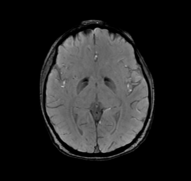
Radiographic features
In general, cystic radiographic changes are appreciated in the basal ganglia and surrounding structures in the brain 3. Generalised cerebral atrophy may also be noted on any imaging modality 3.
CT
CT may reveal regions of hypodensity in the basal ganglia, indicative of cystic degeneration 3.
MRI
MRI is the modality of choice for evaluating patients with neuroferritinopathy 3. Signal changes in the brain include 3:
- T2/FLAIR: hyperintensity in the globus pallidi and putamina, caudate, substantia nigra, and cerebellar nuclei, these are reflective of cystic changes 3
- T1: hypointense ring surrounding aforementioned cystic changes 3
- GRE/SWI: markedly hypointense ring surrounding aforementioned cystic changes 3
Treatment and prognosis
No disease-specific treatment is available and symptomatic management is recommended 2.


 Unable to process the form. Check for errors and try again.