
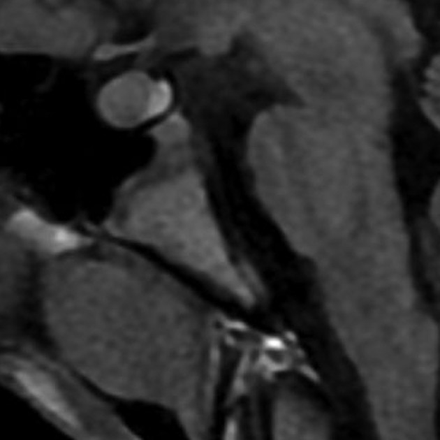
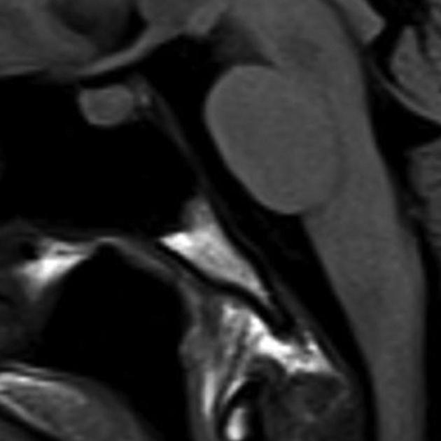
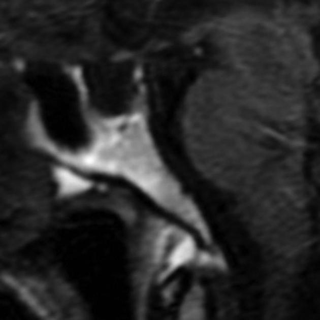
Bone marrow signal of the clivus changes predictably with age and is well assessed with midline non-contrast T1WI without fat saturation. As is seen in the rest of the body the proportion of yellow (fatty) marrow increases with age. Knowledge of these changes allows the diagnosis of pathological processes involving the clivus.
On this page:
Usage
The figures obtained in the study by Kimura et al. have not been precisely replicated in other studies 2,3, and the grading proposed is not widely used in clinical practice. Nonetheless, it is a helpful guide when examining brain MRI and relating bone marrow signal to the patient's age.
Clival bone marrow signal signal varies according to the age of patients 1:
-
patients in their 20s
grade 1: 33%
grade 2: 33%
grade 3: 34%
-
patients in their 50s
grade 1: 0%
grade 2: 40%
grade 3: 60%
-
patients in their 80s
grade 1: 0%
grade 2: 0%
grade 3: 100%
To make things simple, the calvarial bone marrow signal intensity should homogeneously match the white matter on non-contrast T1WI (not fat saturated) as an internal reference standard 3. Most importantly, a finding of low signal in elderly patients warrants comment and review of additional imaging and clinical information.
Classification
Kimura et al. 1 divided the appearance into three grades by comparing the clivus bone marrow to the pons and subcutaneous fat. A high signal was similar to subcutaneous fat, whereas low was iso- to slightly hypointense to the pons.
grade 1: predominantly low signal (>50% of clivus)
grade 2: mixed-signal (low signal 20-50% of clivus)
grade 3: predominantly high signal (>80% of clivus)


 Unable to process the form. Check for errors and try again.