
Nose
Citation, DOI, disclosures and article data
At the time the article was created Henry Knipe had no recorded disclosures.
View Henry Knipe's current disclosuresAt the time the article was last revised Rohit Sharma had no financial relationships to ineligible companies to disclose.
View Rohit Sharma's current disclosures- External nose
- Noses
- External noses
The nose, sometimes referred to as the external nose, is a feature of the face and is composed of soft tissues that extend externally from the skull. It is continuous posteriorly with the nasal cavity. The anterior (piriform) aperture is bounded above by the nasal bones and elsewhere by the two maxillae.
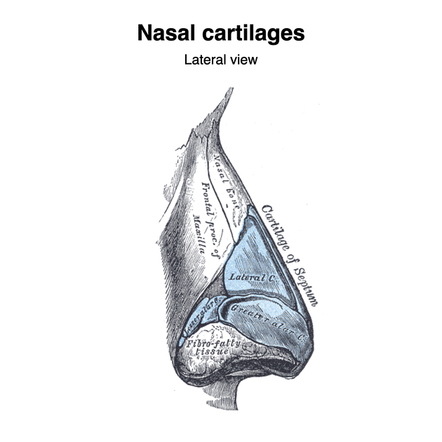
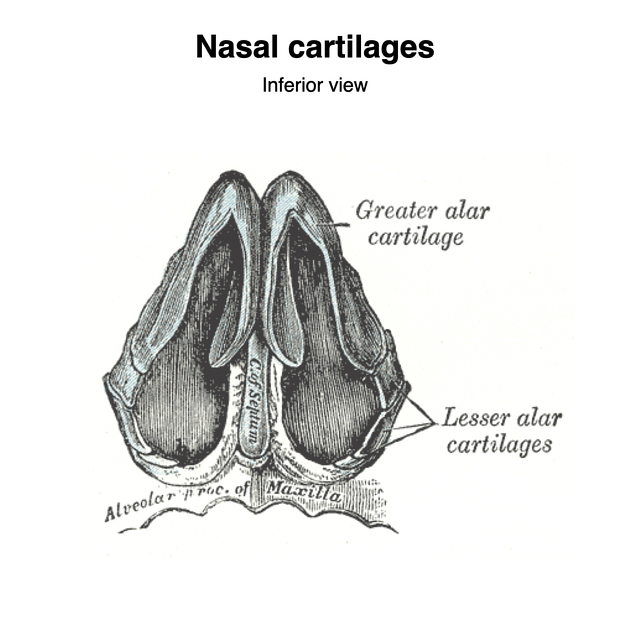
The external nose is formed by nasal bones (bridge of nose), lateral (upper) and greater (alar or lower) nasal cartilages and supported in the midline by the nasal septum. The mucocutaneous area of the nose lies beyond the hair-bearing area 4.
The anterior nares (or nostrils) form the entrance to the nose.
On this page:
Arterial supply
with anastomoses from the supraorbital and supratrochlear arteries (from internal carotid artery) 3
Venous drainage
valveless venous anastomoses with orbital and ophthalmic veins (and thus the cavernous sinus) and the pterygopalatine venous plexus, making this a potential route of infectious spread 3
Lymphatic drainage
receives lymphatics from the anterior nasal cavity and then in lymphatics that accompany the facial vein to the submandibular lymph nodes 2
Innervation
skin of the nose is supplied by the external nasal nerve (ophthalmic division of trigeminal nerve) 4
References
- 1. Anatomy of Orofacial Structures - Enhanced 7th Edition: A Comprehensive Approach, 7e (Anatomy of Orofacial Structures (Brand)). Mosby. ISBN:0323227848. Read it at Google Books - Find it at Amazon
- 2. Lang J. Clinical anatomy of the nose, nasal cavity and paranasal sinuses. Thieme. ISBN:0865773300. Read it at Google Books - Find it at Amazon
- 3. Essentials of otolaryngology. LWW. ISBN:0781747074. Read it at Google Books - Find it at Amazon
- 4. Mcminn. Last's Anatomy. Elsevier Australia. (2003) ISBN:0729537528. Read it at Google Books - Find it at Amazon
Incoming Links
- Nasal septal cartilage
- Organomegaly
- Valsalva manoeuvre
- Pterygopalatine ganglion
- Trichorhinophalangeal syndrome type II
- Superior nasal concha
- Dorsal nasal artery
- Nasal cavity
- Chronic maxillary atelectasis
- Angular artery (facial artery branch)
- Anterior naris
- Nasal cartilages
- Depressor septi nasalis muscle
- Naso-orbitoethmoid (NOE) complex fracture
- Supreme nasal concha
- Nasal septum
- Retropharyngeal space
- Deviated nasal septum
- Rhinolith
- Middle nasal concha
Related articles: Anatomy: Head and neck
- skeleton of the head and neck
-
cranial vault
- scalp (mnemonic)
- fontanelle
-
sutures
- calvarial
- facial
- frontozygomatic suture
- frontomaxillary suture
- frontolacrimal suture
- frontonasal suture
- temporozygomatic suture
- zygomaticomaxillary suture
- parietotemporal suture (parietomastoid suture)
- occipitotemporal suture (occipitomastoid suture)
- sphenofrontal suture
- sphenozygomatic suture
- spheno-occipital suture (not a true suture)
- lacrimomaxillary suture
- nasomaxillary suture
- internasal suture
- basal/internal
- skull landmarks
- frontal bone
- temporal bone
- parietal bone
- occipital bone
- skull base (foramina)
-
facial bones
- midline single bones
- paired bilateral bones
- cervical spine
- hyoid bone
- laryngeal cartilages
-
cranial vault
- muscles of the head and neck
- muscles of the tongue (mnemonic)
- muscles of mastication
-
facial muscles
- epicranius muscle
- circumorbital and palpebral muscles
- nasal muscles
-
buccolabial muscles
- elevators, retractors and evertors of the upper lip
- levator labii superioris alaeque nasalis muscle
- levator labii superioris muscle
- zygomaticus major muscle
- zygomaticus minor muscle
- levator anguli oris muscle
- malaris muscle
- risorius muscle
- depressors, retractors and evertors of the lower lip
- depressor labii inferioris muscle
- depressor anguli oris muscle
- mentalis muscle
- compound sphincter
-
orbicularis oris muscle
- incisivus labii superioris muscle
- incisivus labii inferioris muscle
-
orbicularis oris muscle
- muscle of mastication
- modiolus
- elevators, retractors and evertors of the upper lip
- muscles of the middle ear
- orbital muscles
- muscles of the soft palate
- pharyngeal muscles
- suprahyoid muscles
- infrahyoid muscles
- intrinsic muscles of the larynx
- muscles of the neck
- platysma muscle
- longus colli muscle
- longus capitis muscle
- scalenus anterior muscle
- scalenus medius muscle
- scalenus posterior muscle
- scalenus pleuralis muscle
- sternocleidomastoid muscle
-
suboccipital muscles
- rectus capitis posterior major muscle
- rectus capitis posterior minor muscle
- obliquus capitis superior muscle
- obliquus capitis inferior muscle
- accessory muscles of the neck
- deep cervical fascia
-
deep spaces of the neck
- anterior cervical space
- buccal space
- carotid space
- danger space
- deep cervical fascia
- infratemporal fossa
- masticator space
- parapharyngeal space
- stylomandibular tunnel
- parotid space
- pharyngeal (superficial) mucosal space
- perivertebral space
- posterior cervical space
- pterygopalatine fossa
- retropharyngeal space
- suprasternal space (of Burns)
- visceral space
- surgical triangles of the neck
- orbit
- ear
- paranasal sinuses
- upper respiratory tract
- viscera of the neck
- blood supply of the head and neck
-
arterial supply
-
common carotid artery
- carotid body
- carotid bifurcation
- subclavian artery
- variants
-
common carotid artery
- venous drainage
-
arterial supply
- innervation of the head and neck
-
cranial nerves
- olfactory nerve (CN I)
- optic nerve (CN II)
- oculomotor nerve (CN III)
- trochlear nerve (CN IV)
-
trigeminal nerve (CN V) (mnemonic)
- trigeminal ganglion
- ophthalmic division
- maxillary division
- mandibular division
- abducens nerve (CN VI)
- facial nerve (CN VII)
-
vestibulocochlear nerve (CN VIII)
- vestibular ganglion (Scarpa's ganglion)
- glossopharyngeal nerve (CN IX)
- vagus nerve (CN X)
- (spinal) accessory nerve (CN XI)
- hypoglossal nerve (CN XII)
- parasympathetic ganglia of the head and neck
- cervical sympathetic ganglia
- greater occipital nerve
- third occipital nerve
-
cervical plexus
- muscular branches
- longus capitis
- longus colli
- scalenes
- geniohyoid
- thyrohyoid
-
ansa cervicalis
- omohyoid (superior and inferior bellies separately)
- sternothyroid
- sternohyoid
- phrenic nerve
- contribution to the accessory nerve (CN XI)
- cutaneous branches
- muscular branches
- brachial plexus
- pharyngeal plexus
-
cranial nerves
- lymphatic drainage of the head and neck
- embryological development of the head and neck


 Unable to process the form. Check for errors and try again.