
Nuchal fold
Citation, DOI, disclosures and article data
At the time the article was created Frank Gaillard had no recorded disclosures.
View Frank Gaillard's current disclosuresAt the time the article was last revised Arlene Campos had no financial relationships to ineligible companies to disclose.
View Arlene Campos's current disclosures- Nuchal thickness
- Thickened nuchal fold
- Thick nuchal fold
- Nuchal thickening
- Nuchal fold thickening
The nuchal fold is a normal fold of skin seen at the back of the fetal neck during the second trimester of pregnancy. Increased thickness of the nuchal fold is a soft marker associated with multiple fetal anomalies, and is measured on a routine second trimester ultrasound.
On this page:
Terminology
It should not be confused with nuchal translucency, which is measured in the first trimester.
Epidemiology
Associations
The predominant reason for measuring the nuchal fold is that it is a soft marker for aneuploidy. As an isolated finding, it has a likelihood ratio of 3.25 for Down syndrome 9.
Other associations include:
normal variant (rare <1%)
Pathology
The proposed aetiology of increased nuchal thickness is the result of hydrops or lymphatic obstruction.
ADVERTISEMENT: Supporters see fewer/no ads
Radiographic features
Ultrasound
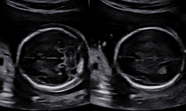
Nuchal fold thickness of >6 mm is abnormal on a routine morphology ultrasound performed at 18-22 weeks.
The nuchal fold is known to increase throughout the second trimester in a normal pregnancy, and may be measured during a broader window of 14 and 24 weeks when required. There is some controversy regarding the normative thresholds at the earlier and later gestations; some authors advocate the use of a nomogram 10, while others suggest that the 6 mm upper limit may be appropriate up to 24 weeks 11.
Technical considerations
Nuchal fold thickness is measured on an axial section through the head at the level of the thalami, cavum septi pellucidi, and cerebellar hemispheres (i.e. in the same plane that is used to assess the posterior fossa structures). One calliper should be placed on the outer edge of the skin, and the other against the outer edge of the occipital bone. The ideal angle of insonation is approximately 30o to the horizontal. This plane is less likely to produce a false positive thickened nuchal fold.
ADVERTISEMENT: Supporters see fewer/no ads
Treatment and prognosis
An abnormally thickened nuchal fold or even a cystic hygroma may resolve, especially toward the third trimester; however, the risk of karyotypic abnormalities is not reduced.
History and etymology
The Terminologia Anatomica refers to the neck as a whole as the "collum". In official Latin anatomical nomenclature, "cervix" refers to the front of the neck and "nucha" to the back (nape) of the neck 12.
See also
Quiz questions
References
- 1. Locatelli A, Piccoli M, Vergani P et al. Critical Appraisal of the Use of Nuchal Fold Thickness Measurements for the Prediction of Down Syndrome. Am J Obstet Gynecol. 2000;182(1 Pt 1):192-7. doi:10.1016/s0002-9378(00)70512-6 - Pubmed
- 2. Ralph Weissleder. Primer of Diagnostic Imaging. (2007) ISBN: 9780323040686 - Google Books
- 3. Vintzileos AM, Egan JF. Adjusting the risk for trisomy 21 on the basis of second-trimester ultrasonography. Am. J. Obstet. Gynecol. 1995;172 (3): 837-44. Am. J. Obstet. Gynecol. (link) - Pubmed citation
- 4. Edward I. Bluth. Ultrasound. (2000) ISBN: 9780865778610 - Google Books
- 5. Maymon R, Sharony R, Grinshpun-cohen J et-al. The best marker combination using the integrated screening test approach for detecting various chromosomal aneuploidies. J Perinat Med. 2005;33 (5): 392-8. doi:10.1515/JPM.2005.071 - Pubmed citation
- 6. Odibo A, Sehdev H, Gerkowicz S, Stamilio D, Macones G. Comparison of the Efficiency of Second-Trimester Nasal Bone Hypoplasia and Increased Nuchal Fold in Down Syndrome Screening. Am J Obstet Gynecol. 2008;199(3):281.e1-5. doi:10.1016/j.ajog.2008.06.078 - Pubmed
- 7. Wolfgang Dähnert. Radiology Review Manual. (2011) ISBN: 9781609139438 - Google Books
- 8. Cho J, Kim K, Lee Y, Toi A. Measurement of Nuchal Skin Fold Thickness in the Second Trimester: Influence of Imaging Angle and Fetal Presentation. Ultrasound Obstet Gynecol. 2005;25(3):253-7. doi:10.1002/uog.1847 - Pubmed
- 9. Agathokleous M, Chaveeva P, Poon LC, Kosinski P, Nicolaides KH. Meta-analysis of second-trimester markers for trisomy 21. (2013) Ultrasound in obstetrics & gynecology : the official journal of the International Society of Ultrasound in Obstetrics and Gynecology. 41 (3): 247-61. doi:10.1002/uog.12364 - Pubmed
- 10. Goynumer G, Arisoy R, Turkmen O, Yayla M. Fetal nuchal skin-fold thickness during the 2nd trimester of pregnancy. (2015) Journal of obstetrics and gynaecology : the journal of the Institute of Obstetrics and Gynaecology. 35 (2): 111-4. doi:10.3109/01443615.2014.937681 - Pubmed
- 11. Singh C & Biswas A. Impact of Gestational Age on Nuchal Fold Thickness in the Second Trimester. J Ultrasound Med. 2014;33(4):687-90. doi:10.7863/ultra.33.4.687 - Pubmed
- 12. FIPAT. Terminologia Anatomica. 2nd Ed. FIPAT.library.dal.ca. Federative International Programme for Anatomical Terminology, 2019. https://fipat.library.dal.ca/TA2/
Incoming Links
Related articles: Pathology: Genitourinary
- obstetrics
-
first trimester
- ultrasound findings in early pregnancy
- embryo/fetus
- beta-hCG levels
- confirming intrauterine gestation
- pregnancy of unknown location (PUL)
- first trimester vaginal bleeding
- early structural scan
- aneuploidy testing
-
second trimester
- fetal biometry
- amniotic fluid volume
- fetal morphology assessment
- soft markers
- amnioreduction
- Doppler ultrasound
- nuchal translucency
- 11-13 weeks antenatal scan
- chorionic villus sampling (CVS) and amniocentesis
- other
- placenta
- placental anatomy
- placental developmental abnormalities
- placenta praevia
- spectrum of abnormal placental villous adherence
- abnormalities of cord insertion
- abruptio placentae
- placental pathology
- vascular pathologies of placenta
- placental infections
- placental masses
- molar pregnancy
- twin placenta
- miscellaneous
-
first trimester
- gynaecology
- acute pelvic pain
- chronic pelvic pain
- uterus
- ovaries
- ovarian follicle
- ovarian torsion
- pelvic inflammatory disease
- ovarian cysts and masses
- paraovarian cyst
- polycystic ovaries
- ovarian hyperstimulation syndrome
- post-hysterectomy ovary
- cervix
- fallopian tube
- other
- male genital tract
- prostate gland
- transrectal ultrasound
- prostate tumours
- infections of the prostate
-
prostatitis
- acute bacterial prostatitis
-
chronic prostatitis
- chronic bacterial prostatitis
- chronic prostatitis and chronic pelvic pain syndrome (CPPS)
- asymptomatic inflammatory prostatitis
- granulomatous prostatitis
- emphysematous prostatitis
- prostatic abscess
-
prostatitis
- benign prostatic hypertrophy
- cystic lesions of the prostate
- prostatic calcification
- prostatic infarction
- testes
-
unilateral testicular lesion
- testicular torsion
- orchitis
- testicular trauma
-
germ cell tumours of the testis
- testicular seminoma
-
non seminomatous germ cell tumours
- mixed germ cell tumour
- yolk sac tumour (endodermal sinus tumour)
- embryonal cell carcinoma
- choriocarcinoma
- testicular teratoma
- testicular epidermoid (teratoma with ectodermal elements only)
- burned out testis tumour
- sex cord / stromal tumours of the testis
- testicular cyst
- testicular lymphoma
- bilateral testicular lesion
- paratesticular lesions
- epididymis
- other
- polyorchidism
- cryptorchidism
- tubular ectasia of the rete testis
- cystadenoma of the rete testis
- testicular sarcoidosis
- testicular tuberculosis
- spermatic cord
- fibrous pseudotumour of the scrotum
- scrotal leiomyosarcoma
- testicular adrenal rest tumours (TARTs)
- tunica vaginalis testis mesothelioma
- splenogonadal fusion
- testicular vasculitis
- abnormal testicular Doppler flow (differential)
-
unilateral testicular lesion
- penis
- prostate gland
- KUB
- kidneys
- normal renal anatomy
- hydronephrosis
- urolithiasis
- renal masses
- renal cystic disease
- renal infection
- vascular
- trauma
- ureter
- normal ureter anatomy
- ureteral stricture
- ureteral dilatation
- ureteral anomalies
- ureteral tumours
- ureteral trauma
- other
- bladder
- kidneys


 Unable to process the form. Check for errors and try again.