
Oesophageal lipomas (or lipomata) are rare fat-containing oesophageal lesions.
On this page:
Epidemiology
They may account for approximately 0.4% of the benign tumours of the alimentary tract 1. There may be greater male predilection. The average age of presentation is around 50 years.
Clinical presentation
They are usually small, asymptomatic and incidentally detected. When large (>3 cm), patients may present with symptoms such as dysphagia, regurgitation, odynophagia, epigastralgia or haemorrhage.
Pathology
As with all lipomas, they are composed entirely of mature adipocytes with or without mesenchymal tissue elements. There exists a variety of histological subtypes which include spindle cell lipoma, angiolipoma, myolipoma, fibrolipoma, myxoid lipoma, and the common classic lipoma. Some lesions may be pedunculated.
Location
In terms of location, they most commonly occur in the upper one-third of the thoracic oesophagus but can potentially occur anywhere from the pharynx to the distal oesophagus.
Radiographic features
Fluoroscopy (barium swallow)
They can appear as smooth intraluminal filling defects. Some may be pedunculated.
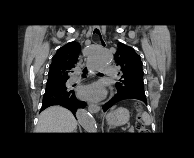
CT
CT characteristically demonstrates a homogeneous fat attenuation in typical lesions.
MRI
Follows fat signal with a high T1 weighted intensity that becomes low intensity on fat-suppressed images.
Differential diagnosis
Possible differential considerations include
- liposarcoma arising from the oesophageal region: it can be usually differentiated from liposarcoma which appears as a heterogeneous lesion with internal septations and soft tissue components without fat attenuation.
For atypical lesions consider other oesophageal lesions such as


 Unable to process the form. Check for errors and try again.