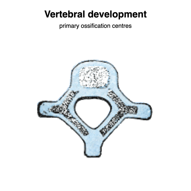
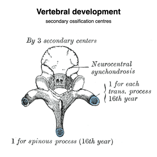
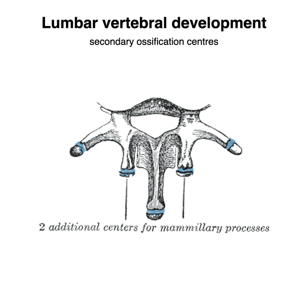
Ossification of the vertebral column is complex but an overview of primary and secondary ossification centers is given below:
Primary ossification centers
The C3-L5 vertebrae typically have three primary ossification centers that start appearing at 9 weeks in utero and finish primary ossification by one year 1-4:
one in the centrum (for most of the vertebral body)
one for each half of the vertebral arch (two in total)
There are some differences for C1 and C2 1-3:
-
C1 (atlas): three primary ossification centers in total
one for the anterior arch
one for each side of the posterior arch (two in total)
-
C2 (axis): five primary ossification centers in total
as for typical vertebrae but has two extra primary ossification centers for the dens (odontoid process)
The primary ossification centers first appear at the cervicothoracic junction at 9 weeks in utero and are followed by upper cervical then thoracolumbar vertebrae with the primary ossification centers of the lumbar vertebral arches the last to appear at approximately 14 weeks in utero 3.
Secondary ossification centers
For the C3-L5 vertebrae there are five secondary ossification centers that appear at puberty and fuse by 25-30 years 1-4:
one at the tip of the spinous process
one at the tip of each transverse process (two in total)
two as ring (or annular) epiphyses at the upper and lower surfaces of the vertebral bodies
C1 and C2 are atypical in that they have some additional or no secondary ossification centers:
C1 (atlas): no secondary ossification centers
-
C2 (axis)
tip of the dens: if it fails to fuse a persistent ossiculum terminale is present


