Citation, DOI, disclosures and article data
Citation:
Weerakkody Y, Knipe H, Feger J, et al. Ossifying fibromyxoid tumor. Reference article, Radiopaedia.org (Accessed on 27 Mar 2025) https://doi.org/10.53347/rID-36876
Ossifying fibromyxoid tumors are soft tissue tumors of uncertain lineage.
It typically manifests in adults. There may be a slight increased male predilection.
The clinical presentation can vary depending on location but usually tends to manifest as a slowly growing painless mass.
Its exact etiology is not clear but a schwannian or chondroid origin has sometimes been favored. It comprises of small round cells dispersed in a myxoid matrix and can contain bone, osteoid, and collagen elements.
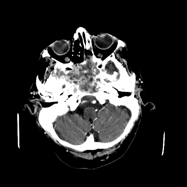
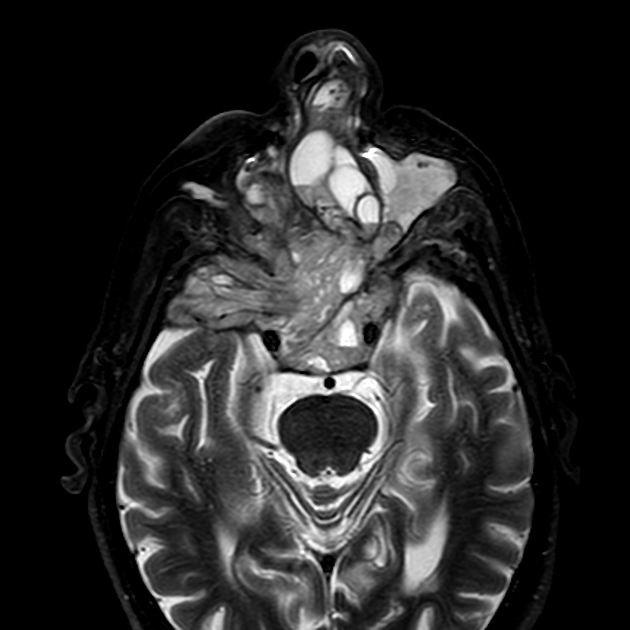
Location
It is reported to most often arise in the subcutaneous extremities. Other reported locations include
-
ribs / chest wall 6
- nasal septum 2
- sinuses
Treatment and prognosis
It is generally considered benign although recurrence following the resection can be common. Complete local resection is considered the best treatment in many cases.
ADVERTISEMENT: Supporters see fewer/no ads
-
1. Ideta S, Nishio J, Aoki M et al. Imaging Findings of Ossifying Fibromyxoid Tumor with Histopathological Correlation: A Case Report. Oncol Lett. 2013;5(4):1301-4. doi:10.3892/ol.2013.1170 - Pubmed
-
2. Blum A, Back W, Naim R, Hörmann K, Riedel F. Ossifying Fibromyxoid Tumor of the Nasal Septum. Auris Nasus Larynx. 2006;33(3):325-7. doi:10.1016/j.anl.2006.01.008 - Pubmed
-
3. Al-Mazrou K, Mansoor A, Payne M, Richardson M. Ossifying Fibromyxoid Tumor of the Ethmoid Sinus in a Newborn: Report of a Case and Literature Review. Int J Pediatr Otorhinolaryngol. 2004;68(2):225-30. doi:10.1016/j.ijporl.2003.09.016 - Pubmed
-
4. Shetty S, Salib R, Nair S, Mathad N, Theaker J. Ossifying Fibromyxoid Tumour of the Sphenoid Sinus. J Laryngol Otol. 2010;124(4):437-40. doi:10.1017/S0022215109991289 - Pubmed
-
5. Cha J, Kwon J, Cho E, Lee C, Yoon Y, Choi S. Ossifying Fibromyxoid Tumor Invading the Spine: A Case Report and Review of the Literature. Skeletal Radiol. 2008;37(12):1137-40. doi:10.1007/s00256-008-0562-0 - Pubmed
-
6. Tateishi U, Gladish G, Kusumoto M et al. Chest Wall Tumors: Radiologic Findings and Pathologic Correlation: Part 1. Benign Tumors. Radiographics. 2003;23(6):1477-90. doi:10.1148/rg.236015526 - Pubmed
-
7. Petscavage-Thomas J, Walker E, Logie C, Clarke L, Duryea D, Murphey M. Soft-Tissue Myxomatous Lesions: Review of Salient Imaging Features with Pathologic Comparison. Radiographics. 2014;34(4):964-80. doi:10.1148/rg.344130110 - Pubmed
-
8. Choi J, Park J, Jin W, Park Y, Ryu K. Sonographic Features of an Ossifying Fibromyxoid Tumor of the Buttock. J Ultrasound Med. 2008;27(5):809-12. doi:10.7863/jum.2008.27.5.809 - Pubmed
Promoted articles (advertising)


 Unable to process the form. Check for errors and try again.