
Osteochondral fracture
Citation, DOI, disclosures and article data
Citation:
Knipe H, Al Kabbani A, Jones J, et al. Osteochondral fracture. Reference article, Radiopaedia.org (Accessed on 28 Mar 2025) https://doi.org/10.53347/rID-63295
rID:
63295
Article created:
Disclosures:
At the time the article was created Henry Knipe had no recorded disclosures.
View Henry Knipe's current disclosures
Last revised:
Disclosures:
At the time the article was last revised Ayla Al Kabbani had no recorded disclosures.
View Ayla Al Kabbani's current disclosures
Revisions:
6 times, by
6 contributors -
see full revision history and disclosures
Systems:
Tags:
Synonyms:
- Osteochondral fractures
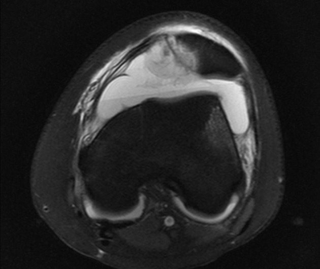
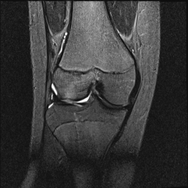
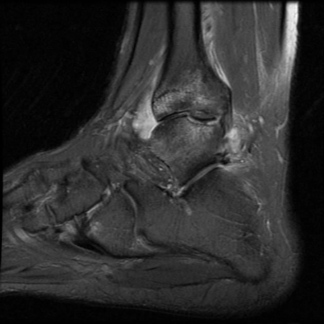
Osteochondral fractures are articular surface injuries involving the articular cartilage and the subchondral bone plate.
Radiographic features
Osteochondral fractures appear as a combination of 1,2:
- fracture line extending to the joint surface
- depression of the subchondral bone plate
- articular surface disruption and/or fragmentation
See also
Quiz questions
error
References
- 1. Gorbachova T, Melenevsky Y, Cohen M, Cerniglia B. Osteochondral Lesions of the Knee: Differentiating the Most Common Entities at MRI. Radiographics. 2018;38(5):1478-95. doi:10.1148/rg.2018180044 [Pubmed]
- 2. Pedersen M, DaCambra M, Jibri Z, Dhillon S, Jen H, Jomha N. Acute Osteochondral Fractures in the Lower Extremities - Approach to Identification and Treatment. Open Orthop J. 2015;9(1):463-74. doi:10.2174/1874325001509010463 [Pubmed]
Incoming Links
Articles:
- Intra-articular fragments
- Osteochondritis dissecans of the knee
- Osteolipoma (soft tissue)
- Snapping hip syndrome
- Subchondral fracture
- Chondral fracture
- Osteochondral fragment
- Osteochondritis dissecans
- Knee radiograph (an approach)
- Intra-articular loose bodies
- Midtarsal sprain
- Triplane fracture
- Osborne-Cotterill lesion
Cases:
- Osteochondritis dissecans of the medial talar dome
- Fracture of an osteochondroma
- Chondral fracture
- Osteochondral knee fracture and Medial patellofemoral ligament injury
- Talus bone osteochondral defect
- Osteochondral fracture - patella
- Osteochondral fracture post lateral patellar dislocation
- Osteochondral knee fracture due to pivot-shift injury
- Chronic osteochondral defect with cystic change
- Osteochondral injury - stage III
- Osteochondral defect
- Osteochondritis dissecans - knee MRI
- Intra-articular loose body - Knee
- Osteochondral injury
- Osteochondral lesion: talus
- Osteochondral injury - talus
- Osteochondral fracture - talus
- Patellar dislocation with chondral injury
- Illustration - displaced osteochondral lesion of the talar dome
- Lateral femoral notch sign - anterior cruciate ligament tear
Multiple choice questions:
Related articles: Fractures
-
fracture
- terminology
- fracture location
- diaphyseal fracture
- metaphyseal fracture
- physeal fracture
- epiphyseal fracture
- fracture types
- avulsion fracture
- articular surface injuries
- complete fracture
- incomplete fracture
- infraction
- compound fracture
- pathological fracture
- stress fracture
- fracture displacement
- fracture location
- fracture healing
- skull fractures
-
facial fractures
- fractures involving a single facial buttress
- alveolar process fractures
- frontal sinus fracture
- isolated zygomatic arch fractures
- mandibular fracture
- nasal bone fracture
- orbital blow-out fracture
- paranasal sinus fractures
- complex fractures
- dental fractures
- fractures involving a single facial buttress
-
spinal fractures
- classification (AO Spine classification systems)
-
cervical spine fracture classification systems
- AO classification of upper cervical injuries
- AO classification of subaxial injuries
- Anderson and D'Alonzo classification (odontoid fracture)
- Roy-Camille classification (odontoid process fracture)
- Gehweiler classifcation (atlas fractures)
- Levine and Edwards classification (hangman fracture)
- Allen and Ferguson classification (subaxial spine injuries)
- subaxial cervical spine injury classification (SLIC)
- thoracolumbar spinal fracture classification systems
- three column concept of spinal fractures (Denis classification)
- classification of sacral fractures
-
cervical spine fracture classification systems
- spinal fractures by region
- spinal fracture types
- classification (AO Spine classification systems)
- rib fractures
- sternal fractures
-
upper limb fractures
- classification
- Rockwood classification (acromioclavicular joint injury)
- AO classification (clavicle fracture)
- Neer classification (clavicle fracture)
- Neer classification (proximal humeral fracture)
- AO classification (proximal humeral fracture)
- AO/OTA classification of distal humeral fractures
- Milch classification (lateral humeral condyle fracture)
- Weiss classification (lateral humeral condyle fracture)
- Bado classification of Monteggia fracture-dislocations (radius-ulna)
- Mason classification (radial head fracture)
- Frykman classification (distal radial fracture)
- Mayo classification (scaphoid fracture)
- Hintermann classification (gamekeeper's thumb)
- Eaton classification (volar plate avulsion injury)
- Keifhaber-Stern classification (volar plate avulsion injury)
- upper limb fractures by region
- shoulder
- clavicular fracture
-
scapular fracture
- acromion fracture
- coracoid process fracture
- glenoid fracture
- humeral head fracture
- proximal humeral fracture
- humeral neck fracture
- arm
- elbow
- forearm
- wrist
-
carpal bones
- scaphoid fracture
- lunate fracture
- capitate fracture
- triquetral fracture
- pisiform fracture
- hamate fracture
- trapezoid fracture
- trapezium fracture
- hand
- shoulder
- classification
- lower limb fractures
- classification by region
- pelvic fractures
- hip fractures
- Pipkin classification (femoral head fracture)
- Garden classification (hip fracture)
- American Academy of Orthopaedic Surgeons classification (periprosthetic hip fracture)
- Cooke and Newman classification (periprosthetic hip fracture)
- Johansson classification (periprosthetic hip fracture)
- Vancouver classification (periprosthetic hip fracture)
- femoral
- knee
- Schatzker classification (tibial plateau fracture)
- AO classification of distal femur fractures
- Meyers and McKeevers classification (anterior cruciate ligament avulsion fracture)
- tibia/fibula
- Watson-Jones classification (tibial tuberosity avulsion fracture)
- ankle
- foot
- Berndt and Harty classification (osteochondral lesions of the talus)
- Sanders CT classification (calcaneal fracture)
- Hawkins classification (talar neck fracture)
- Myerson classification (Lisfranc injury)
- Nunley-Vertullo classification (Lisfranc injury)
- pelvis and lower limb fractures by region
- pelvic fracture
- sacral fracture
- coccygeal fracture
-
hip
- acetabular fracture
- femoral head fracture
-
femoral neck fracture
- subcapital fracture
- transcervical fracture
- basicervical fracture
-
trochanteric fracture
- pertrochanteric fracture
- intertrochanteric fracture
- subtrochanteric fracture
- femur
- mid-shaft fracture
- bisphosphonate-related fracture
- distal femoral fracture
- knee
- avulsion fractures
- Segond fracture
- reverse Segond fracture
- anterior cruciate ligament avulsion fracture
- posterior cruciate ligament avulsion fracture
- arcuate complex avulsion fracture (arcuate sign)
- biceps femoris avulsion fracture
- iliotibial band avulsion fracture
- semimembranosus tendon avulsion fracture
- Stieda fracture (MCL avulsion fracture)
- patellar fracture
- tibial plateau fracture
- avulsion fractures
- leg
- tibial tuberosity avulsion fracture
- tibial shaft fracture
- fibular shaft fracture
- Maisonneuve fracture
- ankle
- foot
- tarsal bones
- metatarsal bones
- phalanges
- classification by region
- terminology


 Unable to process the form. Check for errors and try again.