Citation, DOI, disclosures and article data
Citation:
Weerakkody Y, Walizai T, Campos A, et al. Paraseptal emphysema. Reference article, Radiopaedia.org (Accessed on 30 Mar 2025) https://doi.org/10.53347/rID-17877
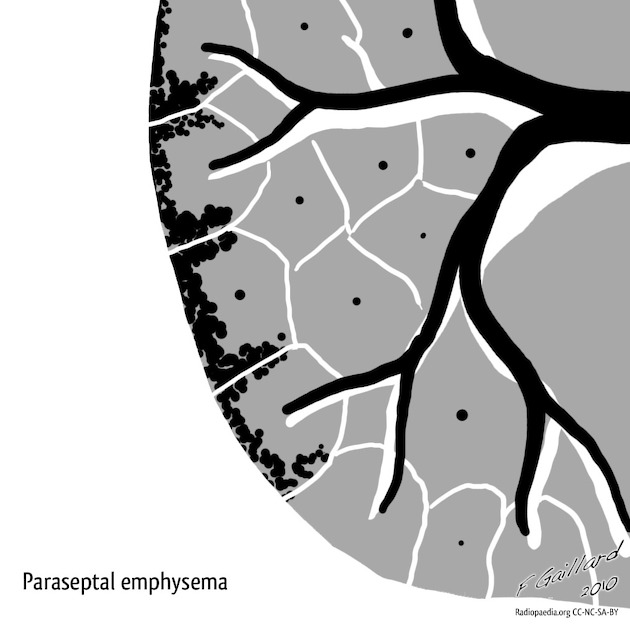
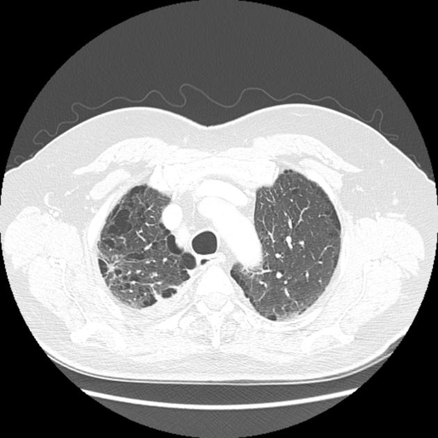
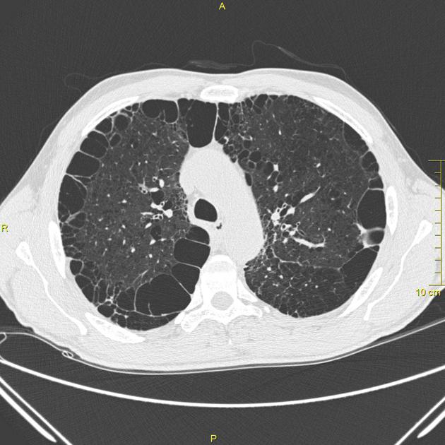
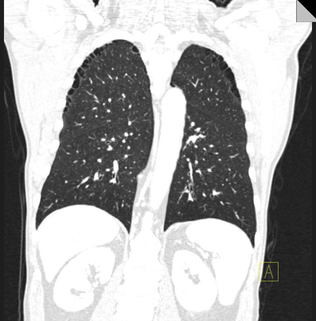
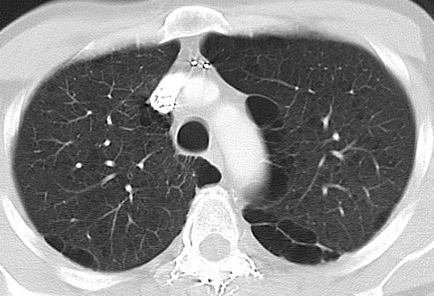
Paraseptal or distal acinar emphysema refers to a morphological subtype of pulmonary emphysema located adjacent to the pleura and septal lines with a peripheral distribution within the secondary pulmonary lobule 1. The affected lobules are almost always subpleural and demonstrate small focal lucencies up to 10 mm in size.
Patients are usually asymptomatic, but the condition is considered to be a cause of pneumothorax in young adults.
Paraseptal emphysema is usually limited in extent occurring most commonly along the dorsal surface of the upper lung and is often associated with fibrosis 4 and may coexist with other types of emphysema.
Aetiology
Smoking and male gender are among risk factors 4.
Associations
Any larger than 10 mm are referred to as subpleural blebs or subpleural bullae.
-
1. Takahashi M, Fukuoka J, Nitta N et al. Imaging of Pulmonary Emphysema: A Pictorial Review. Int J Chron Obstruct Pulmon Dis. 2008;3(2):193-204. doi:10.2147/copd.s2639 - Pubmed
-
2. Swensen S, Aughenbaugh G, Douglas W, Myers J. High-Resolution CT of the Lungs: Findings in Various Pulmonary Diseases. AJR Am J Roentgenol. 1992;158(5):971-9. doi:10.2214/ajr.158.5.1566699 - Pubmed
-
3. Thurlbeck W & Müller N. Emphysema: Definition, Imaging, and Quantification. AJR Am J Roentgenol. 1994;163(5):1017-25. doi:10.2214/ajr.163.5.7976869 - Pubmed
-
4. Araki T, Nishino M, Zazueta O et al. Paraseptal Emphysema: Prevalence and Distribution on CT and Association with Interstitial Lung Abnormalities. Eur J Radiol. 2015;84(7):1413-8. doi:10.1016/j.ejrad.2015.03.010 - Pubmed
-
5. Dyhdalo K & Farver C. Pulmonary Histologic Changes in Marfan Syndrome: A Case Series and Literature Review. Am J Clin Pathol. 2011;136(6):857-63. doi:10.1309/AJCP79SNDHGKQFIN - Pubmed
Multiple choice questions:
Promoted articles (advertising)


 Unable to process the form. Check for errors and try again.