
Pectoralis major injuries are uncommon and include strains, tears and ruptures.
On this page:
Epidemiology
mostly young, physically-active males age 20-40 years old, although has also reported in elderly women 1
associated with weight lifting (mostly bench press), although also reported during various athletic activities e.g. martial arts, boxing, American football, rugby, wrestling, gymnastics, parachuting 1,3,4,6
associated with anabolic steroid use 3, although this is reported inconsistently 1
reported incidence increasing since 1960s 1
Clinical presentation
The diagnosis of pectoralis major injury is often made by history and examination alone 3. Common signs and symptoms are as follows 5:
acute pain and sudden weakness during muscular loading (not always)
swelling/bruising along the chest and/or arm
weakness in shoulder adduction
thinning or loss of the axillary fold (anterior contour between lateral aspect of pectoralis and axilla)
palpable defect
Pathology
The structure of the pectoralis major is complex, with different segments of the two/three muscular heads contributing to shoulder motion in different ways 1,2. Most injuries are thought to occur by indirect mechanism 3, during eccentric loading of the muscle during shoulder abduction and external rotation, such as occurs during lowering of weight during bench press. In this bench press mechanism, the most inferior sternal head fibers are most commonly injured 6.
Location
Injury may occur at several locations:
sternal or clavicular muscle origin
muscle belly: often the result of direct trauma 6
myotendinous junction (~25%) 6
-
tendon
intratendinous
humeral insertion (~60%) 6 +/- bone avulsion
Tears may be either an acute or chronic in nature.
Radiographic features
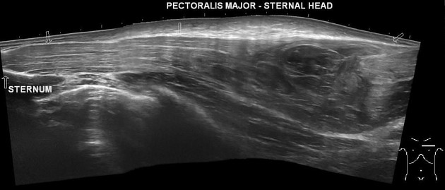
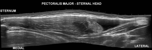
Ultrasound
Ultrasound features that may help diagnose a pectoralis major rupture include 2:
disruption, dehiscence, or absence of tendon distally with retraction of tendon and muscle fibers
associated hemorrhage (initially hypoechoic, becoming progressively heterogeneous with hematoma organization)
abnormal echotexture of muscle belly if muscle belly injury
MRI
A dedicated study of pectoralis muscle may be required and a typical shoulder MRI usually does not allow optimal visualization of the pectoralis major muscle 2. In obtaining images, respiratory motion artifact may be minimized by abdominal breathing techniques.
Features characteristic of injury include:
tendon absence distally with retraction
fluid gap in place of tendon
Radiology report
The following features should be commented on as it aids in classification and treatment for these injuries 6:
-
location
muscle origin/belly
myotendinous junction or distal free tendon
avulsion injury at the enthesis
-
thickness in AP dimension
partial thickness, anterior tendon vs posterior tendon
full-thickness
width in craniocaudal dimension: complete vs incomplete
Treatment and prognosis
Optimal methods for treatment of pectoralis major tears remain under investigation and depends on location and extent of tear, chronicity, and patient factors. There is increasing evidence that surgical management may improve outcome in physically active patients 5.
Complete tears, particularly of the tendon or myotendinous junction, are more commonly managed by surgical repair, consisting of either suturing or bone tunneling techniques. Near-complete recovery of shoulder adduction strength is common 3-5.
Partial ruptures or low-demand patients are often managed non-operatively.


 Unable to process the form. Check for errors and try again.