
Perirenal lymphoceles are the most common cause of perinephric fluid collection. They can occur after transplant in up to 25% of cases.
On this page:
Diagnosis
Ultrasound is the primary method of identification and monitoring of small lymphoceles, which share appearances with seroma, but the latter tend to regress over time. Lymphocoele and seroma can be differentiated from a urine leak by aspiration and assessing creatinine content relative to serum (which would be increased for urine) 6.
Clinical presentation
Perirenal lymphocele is usually asymptomatic, but some can be large enough to cause abdominal discomfort, transplant hydronephrosis and infection, venous thrombosis, and lower limb edema 6,9.
Pathology
Perirenal lymphocele is the most common cause of perinephric fluid collection post-transplant, tending to occur 4-8 weeks after surgery, but can occur years later. Transection and inadequate ligation of iliac lymphatics during the surgery results in lymphatic leakage 6.
Radiographic features
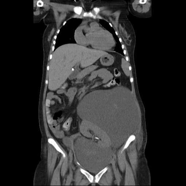
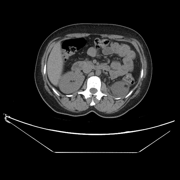
Post-transplant lymphocele often occur between the transplanted kidney and bladder 6, but hilar and superolateral locations are also not uncommon 10.
Ultrasound
Nonspecific anechoic perirenal collection, sometimes with septations. Echogenic debris is possible if they become infected 6.
CT
Round, hypoattenuating collection similar to a seroma, with houdsfield unit values close to water, below that of abscess or hematoma 6. Calcification is rare 5.
MRI
May be helpful to exclude the presence of blood 6.
Treatment and prognosis
Small lymphoceles may be conservatively managed with ultrasound monitoring. Aspiration may be helpful for diagnosis, but lymphocoeles commonly recur, and repeat aspirations risk introduction of infection. Therefore, lymphoceles requiring intervention can be managed with prolonged drainage, sclerotherapy, or surgery (making a peritoneal window for marsupialization) 6-8.
Complications
Perirenal lymphocele can compress adjacent structures and cause transplant hydronephrosis, infection, ipsilateral lower limb venous stasis or impaired lymphatic drainage 6,9.
Differential diagnosis
Imaging differential considerations include:
perirenal seroma - tends to regress over time
urinoma - has high creatinine relative to serum


 Unable to process the form. Check for errors and try again.