Citation, DOI, disclosures and article data
Citation:
Sarangi P, Glick Y, Gaillard F, Piglet sign (osmotic demyelination). Reference article, Radiopaedia.org (Accessed on 23 Mar 2025) https://doi.org/10.53347/rID-63574
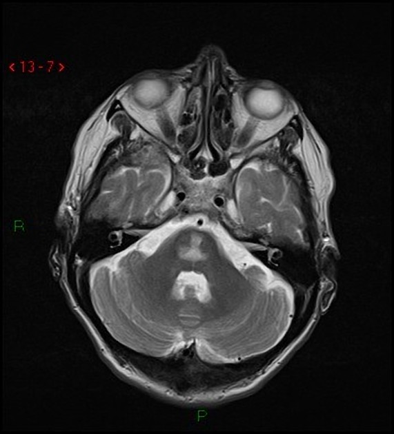
The piglet sign is seen in osmotic demyelination syndrome. It refers to the appearance of the upper pons in axial T2 and FLAIR images. The areas of coalescent T2 signal increase are reminiscent of a pig's snout, with other features on axial MR images resembling the rest of the face of a piglet with the temporal lobes representing ears, the carotid arteries represent the eyes and the fourth ventricle as the mouth 1,2.
The area of abnormally increased T2 signal is perhaps better known as the trident sign.
-
1. Judith Wagner, Stefanie Mueller-Schunk, Christoph Schankin. The Piglet Sign: MRI Findings in Central Pontine Myelinolysis. (2016) Clinical Neuroradiology. 18 (3): 191. doi:10.1007/s00062-008-8025-5
-
2. Beh SC. Temporal evolution of the trident and piglet signs of osmotic demyelination syndrome. (2017) Journal of the neurological sciences. 373: 268-273. doi:10.1016/j.jns.2017.01.024 - Pubmed
Promoted articles (advertising)


 Unable to process the form. Check for errors and try again.