
Pilon fracture (tibia)
Citation, DOI, disclosures and article data
At the time the article was created Yuranga Weerakkody had no recorded disclosures.
View Yuranga Weerakkody's current disclosuresAt the time the article was last revised Arlene Campos had no financial relationships to ineligible companies to disclose.
View Arlene Campos's current disclosures- Pilon fractures
- Pilon fracture of the distal tibia
- Tibial plafond fracture
- Plafond fracture
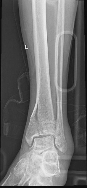
Pilon fractures are a type of distal tibial fracture involving the tibial plafond. These are considered to represent 1-10% of all lower limb fractures 6.
On this page:
Mechanism
Typically, these are high energy injuries and occur as a result of an axial loading which drives the talus into the tibial plafond ref.
Classification
Several classification systems exist. Frequently adopted is the one proposed by Ruedi and Allgower 5:
type I: articular fracture with minimal or no displacement
type II: displacement of the articular surface but with minimal or no comminution
type III: marked comminution as well as articular impaction
Radiographic features
Plain radiograph / CT
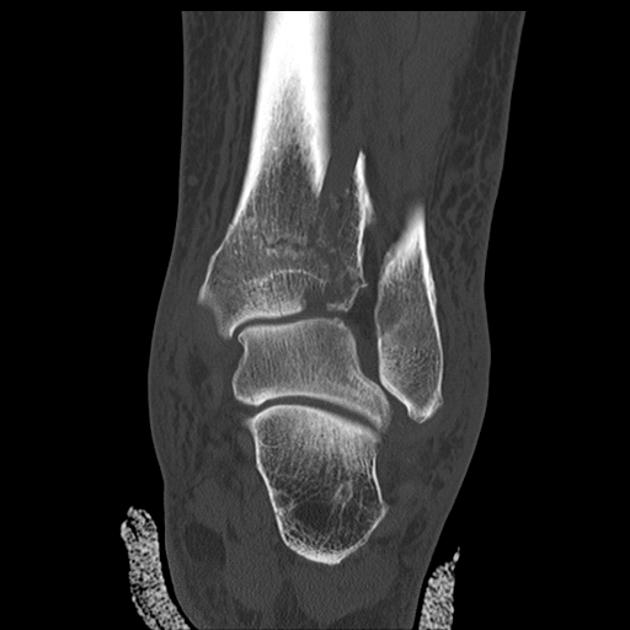
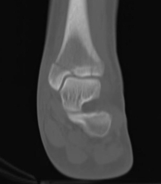
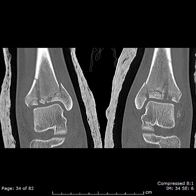
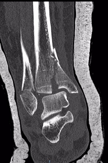
by definition, the fracture involves the tibial plafond and the distal tibial articular surface
associated fractures of the fibula can be evident, with or without ankle dislocation
CT can further define the fracture pattern and assess for tendon subluxation
History and etymology
Derived from the French word pilon (pestle), an instrument used for crushing and pounding and usually used with a mortar.
References
- 1. Cheung Y, Perrich K, Gui J, Koval K, Goodwin D. MRI of Isolated Distal Fibular Fractures with Widened Medial Clear Space on Stressed Radiographs: Which Ligaments Are Interrupted? AJR Am J Roentgenol. 2009;192(1):W7-12. doi:10.2214/AJR.08.1092 - Pubmed
- 2. Topliss CJ, Jackson M, Atkins RM. Anatomy of pilon fractures of the distal tibia. J Bone Joint Surg Br. 2005;87 (5): 692-7. doi:10.1302/0301-620X.87B5.15982 - Pubmed citation
- 3. Haapamaki VV, Kiuru MJ, Koskinen SK. Ankle and foot injuries: analysis of MDCT findings. AJR Am J Roentgenol. 2004;183 (3): 615-22. AJR Am J Roentgenol (full text) - Pubmed citation
- 4. Tscherne, H. 1933-, Schatzker, Joseph.. Major Fractures of the Pilon, the Talus, and the Calcaneus: Current Concepts of Treatment. (1993) ISBN: 3540558373 - Google Books
- 5. Chen S, Wu P, Lee Y. Long-Term Results of Pilon Fractures. Arch Orthop Trauma Surg. 2007;127(1):55-60. doi:10.1007/s00402-006-0225-3 - Pubmed
- 6. Sirkin M & Sanders R. The Treatment of Pilon Fractures. Orthop Clin North Am. 2001;32(1):91-102. doi:10.1016/s0030-5898(05)70196-6 - Pubmed
Incoming Links
Related articles: Fractures
-
fracture
- terminology
- fracture location
- diaphyseal fracture
- metaphyseal fracture
- physeal fracture
- epiphyseal fracture
- fracture types
- avulsion fracture
- articular surface injuries
- complete fracture
- incomplete fracture
- infraction
- compound fracture
- pathological fracture
- stress fracture
- fracture displacement
- fracture location
- fracture healing
- skull fractures
-
facial fractures
- fractures involving a single facial buttress
- alveolar process fractures
- frontal sinus fracture
- isolated zygomatic arch fractures
- mandibular fracture
- nasal bone fracture
- orbital blow-out fracture
- paranasal sinus fractures
- complex fractures
- dental fractures
- fractures involving a single facial buttress
-
spinal fractures
- classification (AO Spine classification systems)
-
cervical spine fracture classification systems
- AO classification of upper cervical injuries
- AO classification of subaxial injuries
- Anderson and D'Alonzo classification (odontoid fracture)
- Roy-Camille classification (odontoid process fracture)
- Gehweiler classifcation (atlas fractures)
- Levine and Edwards classification (hangman fracture)
- Allen and Ferguson classification (subaxial spine injuries)
- subaxial cervical spine injury classification (SLIC)
- thoracolumbar spinal fracture classification systems
- three column concept of spinal fractures (Denis classification)
- classification of sacral fractures
-
cervical spine fracture classification systems
- spinal fractures by region
- spinal fracture types
- classification (AO Spine classification systems)
- rib fractures
- sternal fractures
-
upper limb fractures
- classification
- Rockwood classification (acromioclavicular joint injury)
- AO classification (clavicle fracture)
- Neer classification (clavicle fracture)
- Neer classification (proximal humeral fracture)
- AO classification (proximal humeral fracture)
- AO/OTA classification of distal humeral fractures
- Milch classification (lateral humeral condyle fracture)
- Weiss classification (lateral humeral condyle fracture)
- Bado classification of Monteggia fracture-dislocations (radius-ulna)
- Mason classification (radial head fracture)
- Frykman classification (distal radial fracture)
- Mayo classification (scaphoid fracture)
- Hintermann classification (gamekeeper's thumb)
- Eaton classification (volar plate avulsion injury)
- Keifhaber-Stern classification (volar plate avulsion injury)
- upper limb fractures by region
- shoulder
- clavicular fracture
-
scapular fracture
- acromion fracture
- coracoid process fracture
- glenoid fracture
- humeral head fracture
- proximal humeral fracture
- humeral neck fracture
- arm
- elbow
- forearm
- wrist
-
carpal bones
- scaphoid fracture
- lunate fracture
- capitate fracture
- triquetral fracture
- pisiform fracture
- hamate fracture
- trapezoid fracture
- trapezium fracture
- hand
- shoulder
- classification
- lower limb fractures
- classification by region
- pelvic fractures
- hip fractures
- Pipkin classification (femoral head fracture)
- Garden classification (hip fracture)
- American Academy of Orthopaedic Surgeons classification (periprosthetic hip fracture)
- Cooke and Newman classification (periprosthetic hip fracture)
- Johansson classification (periprosthetic hip fracture)
- Vancouver classification (periprosthetic hip fracture)
- femoral
- knee
- Schatzker classification (tibial plateau fracture)
- AO classification of distal femur fractures
- Meyers and McKeevers classification (anterior cruciate ligament avulsion fracture)
- tibia/fibula
- Watson-Jones classification (tibial tuberosity avulsion fracture)
- ankle
- foot
- Berndt and Harty classification (osteochondral lesions of the talus)
- Sanders CT classification (calcaneal fracture)
- Hawkins classification (talar neck fracture)
- Myerson classification (Lisfranc injury)
- Nunley-Vertullo classification (Lisfranc injury)
- pelvis and lower limb fractures by region
- pelvic fracture
- sacral fracture
- coccygeal fracture
-
hip
- acetabular fracture
- femoral head fracture
-
femoral neck fracture
- subcapital fracture
- transcervical fracture
- basicervical fracture
-
trochanteric fracture
- pertrochanteric fracture
- intertrochanteric fracture
- subtrochanteric fracture
- femur
- mid-shaft fracture
- bisphosphonate-related fracture
- distal femoral fracture
- knee
- avulsion fractures
- Segond fracture
- reverse Segond fracture
- anterior cruciate ligament avulsion fracture
- posterior cruciate ligament avulsion fracture
- arcuate complex avulsion fracture (arcuate sign)
- biceps femoris avulsion fracture
- iliotibial band avulsion fracture
- semimembranosus tendon avulsion fracture
- Stieda fracture (MCL avulsion fracture)
- patellar fracture
- tibial plateau fracture
- avulsion fractures
- leg
- tibial tuberosity avulsion fracture
- tibial shaft fracture
- fibular shaft fracture
- Maisonneuve fracture
- ankle
- foot
- tarsal bones
- metatarsal bones
- phalanges
- classification by region
- terminology


 Unable to process the form. Check for errors and try again.