
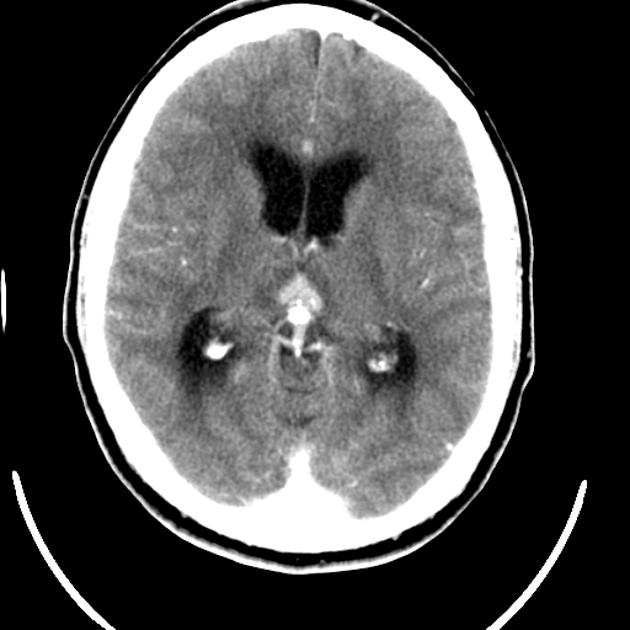
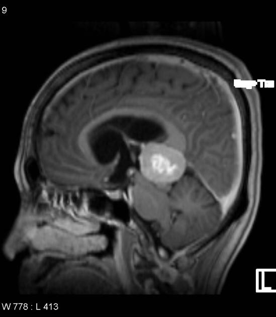
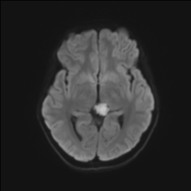
Masses in the pineal region have a relatively broad differential because of the variety of cell types found in the region.
On this page:
Terminology
The term pinealoma was historically used to refer to both pineal parenchymal tumours and germinomas, which are the two most common categories of primary pineal region tumours 6. Germinomas were sometimes called the "two-cell" pattern type of pinealoma, due to the presence of both lymphocytes and neoplastic germ cells that were reminiscent of teratomas 6. The term pinealoma has since fallen in popularity.
Clinical presentation
Many pineal region lesions, especially small ones that do not significantly change in size, such as cysts, are typically discovered incidentally. For larger lesions, patients often present clinically with symptoms related to mass effect. Compression of the tectal plate may cause an upward gaze palsy (Parinaud syndrome). Compression of the cerebral aqueduct may cause obstructive hydrocephalus.
Radiographic features
Intrinsic pineal tissue masses tend to cause upward displacement of the internal cerebral veins. This is in distinction to tentorial meningiomas, which depress the cerebral veins.
Differential diagnosis
pineal cyst (most common benign pineal region mass)
-
pineal germinoma: most common (~50% of primary pineal region tumours)
pineal yolk sac carcinoma (endodermal sinus tumour)
-
pineal parenchymal tumours (~30% of primary pineal region tumours)
glioma (usually tectal in origin, astrocytic in histology) (~5% of primary pineal region tumours)
primary pineal malignant melanoma 5
inclusion cysts (dermoid/epidermoid) 4
meningioma near the pineal region
-
rare vascular lesions
cavernoma in the pineal region
If invasive, a tectal plate mass may be difficult to distinguish from a pineal mass.


 Unable to process the form. Check for errors and try again.