
Presacral space
Citation, DOI, disclosures and article data
At the time the article was created Bruno Di Muzio had no recorded disclosures.
View Bruno Di Muzio's current disclosuresAt the time the article was last revised Zemar Vajuhudeen had no recorded disclosures.
View Zemar Vajuhudeen's current disclosures- Pre-sacral space
- Presacral spaces
- Pre-sacral spaces
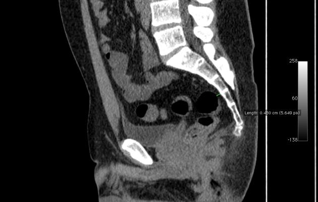
The presacral space is located between the rectum and the sacrococcygeal part of the spine.
On this page:
Gross anatomy
Contents
The presacral space contains a variety of tissue:
- fat
- mesenchymal tissue
- lymph nodes
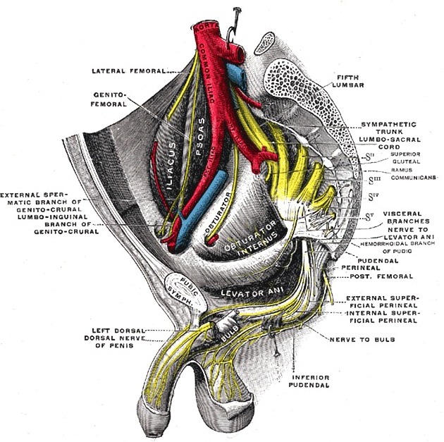
- nerve plexuses
- blood vessels
Boundaries
- superior - peritoneal reflections
- inferior - levator ani and coccygeus muscles
- anterior - rectum (posterior wall)
- posterior - sacrum / coccyx
- laterally - ureter, iliac vessels 1
Radiographic appearance
An increase in the width of the presacral space measurement is considered indicative of pathology in the rectum or other structures in the pelvis. This alteration is usually seen in barium enema studies and on plain film.
The upper limits of normal for the presacral space width is 15 mm in patients younger than 45 years old 2. However, some patients have measurements over 15 mm with no apparent reason (fewer still have 'normal' measurements above 20 mm), which increases with older age. Factors such as sex and weight of the patient should also be taken into consideration 2.
Related pathology
References
- 1. Kocaoglu M, Frush DP. Pediatric presacral masses. Radiographics. 26 (3): 833-57. doi:10.1148/rg.263055102 - Pubmed citation
- 2. Kattan KR, King AY. Presacral space revisited. AJR Am J Roentgenol. 1979;132 (3): 437-9. AJR Am J Roentgenol (abstract) - Pubmed citation
Incoming Links
Related articles: Anatomy: Abdominopelvic
- skeleton of the abdomen and pelvis
- muscles of the abdomen and pelvis
- spaces of the abdomen and pelvis
- anterior abdominal wall
- posterior abdominal wall
- abdominal cavity
- pelvic cavity
- perineum
- abdominal and pelvic viscera
- gastrointestinal tract
- spleen
- hepatobiliary system
-
endocrine system
-
adrenal gland
- adrenal vessels
- chromaffin cells
- variants
- pancreas
- organs of Zuckerkandl
-
adrenal gland
-
urinary system
-
kidney
- renal pelvis
- renal sinus
- avascular plane of Brodel
-
variants
- number
- fusion
- location
- shape
- ureter
- urinary bladder
- urethra
- embryology
-
kidney
- male reproductive system
-
female reproductive system
- vulva
- vagina
- uterus
- adnexa
- Fallopian tubes
- ovaries
- broad ligament (mnemonic)
- variant anatomy
- embryology
- blood supply of the abdomen and pelvis
- arteries
-
abdominal aorta
- inferior phrenic artery
- coeliac artery
- superior mesenteric artery
- middle suprarenal artery
- renal artery (variant anatomy)
- gonadal artery (ovarian artery | testicular artery)
- inferior mesenteric artery
- lumbar arteries
- median sacral artery
-
common iliac artery
- external iliac artery
-
internal iliac artery (mnemonic)
- anterior division
- umbilical artery
- superior vesical artery
- obturator artery
- vaginal artery
- inferior vesical artery
- uterine artery
- middle rectal artery
-
internal pudendal artery
- inferior rectal artery
-
perineal artery
- posterior scrotal artery
- transverse perineal artery
- artery to the bulb
- deep artery of the penis/clitoris
- dorsal artery of the penis/clitoris
- inferior gluteal artery
- posterior division (mnemonic)
- variant anatomy
- anterior division
-
abdominal aorta
- portal venous system
- veins
- anastomoses
- arterioarterial anastomoses
- portal-systemic venous collateral pathways
- watershed areas
- arteries
- lymphatics
- innervation of the abdomen and pelvis
- thoracic splanchnic nerves
- lumbar plexus
-
sacral plexus
- lumbosacral trunk
- sciatic nerve
- superior gluteal nerve
- inferior gluteal nerve
- nerve to piriformis
- perforating cutaneous nerve
- posterior femoral cutaneous nerve
- parasympathetic pelvic splanchnic nerves
- pudendal nerve
- nerve to quadratus femoris and inferior gemellus muscles
- nerve to internal obturator and superior gemellus muscles
- autonomic ganglia and plexuses


 Unable to process the form. Check for errors and try again.