
Pulmonary artery pseudoaneurysm refers to a pseudoaneurysm arising from the pulmonary arteries.
On this page:
Pathology
A pseudoaneurysm results from a tear or disruption of all three layers of the vessel wall. Extravasated blood is contained by compressed extravascular tissue or a clot, which makes up the wall of the pseudoaneurysm. Histologically, a pseudoaneurysm involves only the external layers of the arterial wall (the media and adventitia).
They can arise from a number of causes including:
- infection
- can occur from various infective aetiology including mycotic, syphilis, mycobacterial infection
- associated neoplasms
- trauma - post traumatic pulmonary arterial pseudoaneurysm (usually rare)
- extravascular trauma
- mostly occur from penetrating trauma such as gunshot injuries or stab wounds
- pseudoaneurysms due to blunt trauma have been rarely reported 6
- intravascular trauma: iatrogenic insults: rare cases due to Swan-Ganz catheter placement have been rarely reported 5
- extravascular trauma
- pulmonary valvular and post-valvular stenosis and increased flow due to left-to-right shunting 8
Associations
- conditions that predispose to vascular wall anomalies such as 5
- pulmonary hypertension 8
Location
In general they tend to have a strong predilection for the peripheral pulmonary arteries with most located in segmental and subsegmental pulmonary arteries 1.
Radiographic features
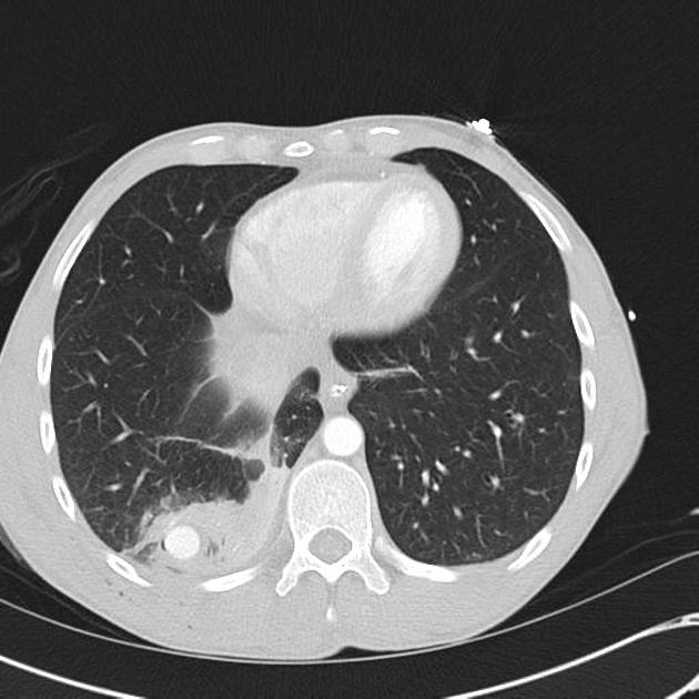
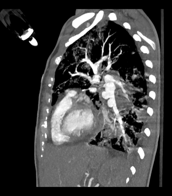
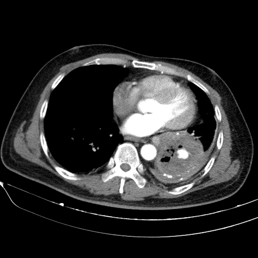
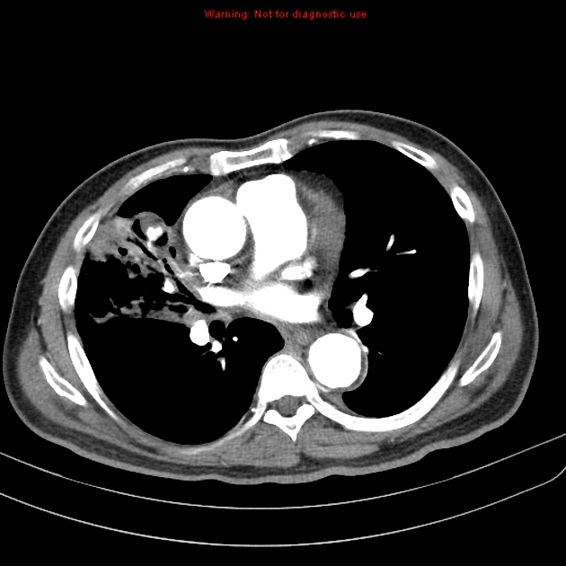
CT chest / CT pulmonary angiography
CT pulmonary angiography is considered the gold standard and may show focal dilatation of a segment of the involved pulmonary arterial segment (they may appear contained or not contained depending on status at time of scan).
Treatment and prognosis
Prognosis is not well described (probably due to rarity) and may depend on multiple variables such as associated injuries/pathology, age, haemorrhagic diathesis, size and delay before diagnosis. Treatment options may include surgical lung resection, embolotherapy / coil embolisation).


 Unable to process the form. Check for errors and try again.