
Radicular veins are small valveless veins that accompany lumbosacral spinal nerve roots and play a role in venous drainage from the spinal cord 1.
On this page:
Gross anatomy
Radicular veins follow the spinal nerve roots and exit the spinal canal through the intervertebral foramina, and as such, can be very long 5. They can puncture the dura with the nerve root or through a separate foramen.
Radicular veins form part of the venous drainage pathway for the conus medullaris and cauda equina 6, draining venous blood to the internal vertebral venous plexus and onto the systemic circulation 8.
A few types of radicular veins have been described 6:
great radicular veins: few; 0.5-1.2 mm, around the thoracolumbar junction
accessory great radicular veins: common; accompany a lumbosacral nerve root or filum terminale
small radicular veins: numerous; microscopic; embedded in endoneurium
As they lack valves, radicular veins can help equalise pressure across different venous territories, which may protect the spinal cord under certain conditions. In normal conditions, four structures can act as anti-reflux devices without completely preventing venous reflux 1:
valve-like dural folds
meandering configuration and widening of the radicular veins
narrowing of the dural part of the radicular veins
venous mural smooth muscle fibres
Radiographic features
MRI
Time-resolved angiographic sequences (e.g. TWIST) may be the only reliable way to differentiate spinal arteries and veins 5.
Signal characteristics
T2: low signal 5
T1C+: high signal 5
DSA (angiography)
gold standard for identifying spinal vascular malformations
helps locate feeding arteries and draining radicular veins
Clinical importance
spinal dural arteriovenous fistulas (dAVFs): abnormal connections between arteries and veins near the dura, often involving radicular veins 3
impaired radicular vein drainage can cause venous hypertension, spinal cord swelling, and spinal cord ischaemia ref
pathway for tumour spread: radicular veins connect to the Batson venous plexus through which tumour cells (especially prostate, breast, and lung cancers) can spread to the vertebral column and brain; this explains why spinal metastases are common even without direct arterial involvement 7
risk in spinal surgery and interventions: as radicular veins are small and fragile, they can be inadvertently damaged during spinal surgery or interventions like lumbar punctures, which can lead to venous bleeding, spinal haematomas, or spinal cord malperfusion ref
Practical points
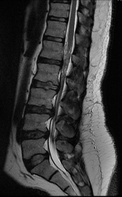
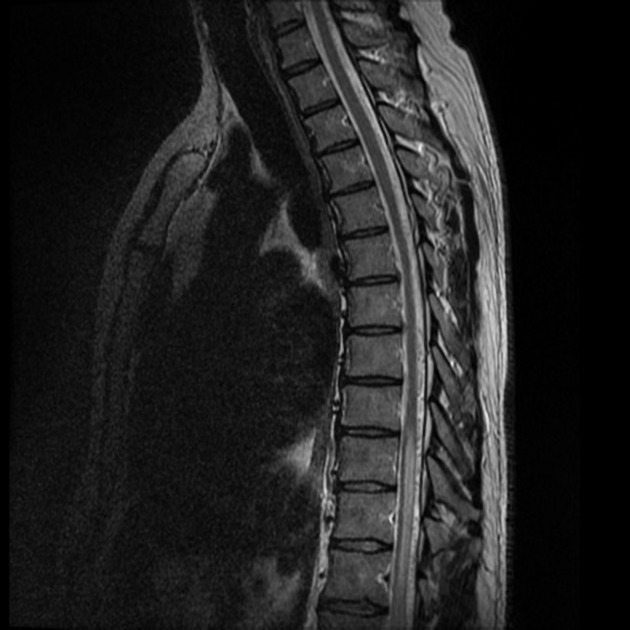
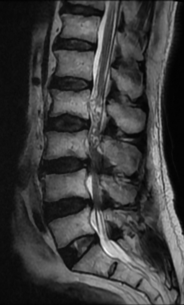
enhancing radicular veins, which is a non-pathological finding, can be mistaken for as enhancing nerve roots but can be differentiated if seen to joint with radiculomedullary veins on the surface of the conus medullaris 2,6
congested coiled radicular veins about the conus medullaris can be misdiagnosed as spinal dural arteriovenous fistulas or redundant cauda equina nerve roots, especially if there is spinal canal stenosis


 Unable to process the form. Check for errors and try again.