Retrobulbar haemorrhage
Updates to Article Attributes
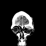
Retrobulbar haemorrhage is the presence of a post septal orbital haematoma and is usually due to craniofacial trauma causing an extraconal haematoma. It may cause orbital compartment syndrome which is an ophthalmologic emergency.
Clinical features
If small, there may be no orbital or ocular symptoms. If large enough to create mass effect on the contents of the orbit (particularly the optic nerve) and orbital compartment syndrome, features include:
- visual disturbance
- proptosis
- pain
- orbital bruising and swelling
- features of head and/or facial trauma
Etiology
The most common cause is trauma, almost always secondary to orbital fractures. Other causes include:
- orbital surgery
- other surgery e.g. sinus or neurosurgical procedures
- haemorrhage from an intraorbital vascular mass e.g. orbital vascular malformation or metastases
- coagulopathies
Radiographic features
CT is the primary imaging modality for cranio-orbital trauma. The orbit is best assessed in bone and soft tissue coronal reconstructions.
Extraconal haematoma has a classic appearance of a confined lentiform hyperdense haematoma, analagous to an intracranial extradural haemorrhage. It is usually located in the superior half of the orbit and almost always adjacent to a fracture of the bony orbit. If large enough they can exert mass effect on the contents of the orbital, causing stretching of the optic nerve and proptosis.
Extraconal haematomas can be difficult to distinguish from a subperiosteal haematoma.
Treatment
Orbital compartment syndrome is a ophthalmologicalan ophthalmologic emergency and requires urgent lateral canthotomy and cantholysis to decompress the orbit and preserve ocular function.
-<p><strong>Retrobulbar haemorrhage</strong> is the presence of a post septal orbital haematoma and is usually due to craniofacial trauma causing an extraconal haematoma. </p><h4>Clinical features</h4><p>If small, there may be no orbital or ocular symptoms. If large enough to create mass effect on the contents of the orbit (particularly the optic nerve) and <a title="Orbital compartment syndrome" href="/articles/orbital-compartment-syndrome">orbital compartment syndrome</a>, features include:</p><ul>- +<p><strong>Retrobulbar haemorrhage</strong> is the presence of a post septal orbital haematoma and is usually due to craniofacial trauma causing an <strong>extraconal haematoma</strong>. It may cause orbital compartment syndrome which is an ophthalmologic emergency.</p><h4>Clinical features</h4><p>If small, there may be no orbital or ocular symptoms. If large enough to create mass effect on the contents of the orbit (particularly the optic nerve) and <a href="/articles/orbital-compartment-syndrome">orbital compartment syndrome</a>, features include:</p><ul>
-<li><a title="Proptosis" href="/articles/proptosis-1">proptosis</a></li>- +<li><a href="/articles/proptosis-1">proptosis</a></li>
-</ul><h4>Radiographic features</h4><p>CT is the primary imaging modality for cranio-orbital trauma. The orbit is best assessed in bone and soft tissue coronal reconstructions.</p><p>Extraconal haematoma has a classic appearance of a confined lentiform hyperdense haematoma, analagous to an intracranial extradural haemorrhage. It is usually located in the superior half of the orbit and almost always adjacent to a fracture of the bony orbit. If large enough they can exert mass effect on the contents of the orbital, causing stretching of the optic nerve and proptosis. </p><p>Extraconal haematomas can be difficult to distinguish from a subperiosteal haematoma.</p><h4>Treatment</h4><p>Orbital compartment syndrome is a ophthalmological emergency and requires urgent lateral canthotomy and cantholysis to decompress the orbit and preserve ocular function.</p>- +</ul><h4>Radiographic features</h4><p>CT is the primary imaging modality for cranio-orbital trauma. The orbit is best assessed in bone and soft tissue coronal reconstructions.</p><p>Extraconal haematoma has a classic appearance of a confined lentiform hyperdense haematoma, analagous to an intracranial extradural haemorrhage. It is usually located in the superior half of the orbit and almost always adjacent to a fracture of the bony orbit. If large enough they can exert mass effect on the contents of the orbital, causing stretching of the optic nerve and proptosis. </p><p>Extraconal haematomas can be difficult to distinguish from a subperiosteal haematoma.</p><h4>Treatment</h4><p>Orbital compartment syndrome is an ophthalmologic emergency and requires urgent lateral canthotomy and cantholysis to decompress the orbit and preserve ocular function.</p>
References changed:
- 1. McCallum E, Keren S, Lapira M, Norris JH. Orbital Compartment Syndrome: An Update With Review Of The Literature. (2019) Clinical ophthalmology (Auckland, N.Z.). 13: 2189-2194. <a href="https://doi.org/10.2147/OPTH.S180058">doi:10.2147/OPTH.S180058</a> - <a href="https://www.ncbi.nlm.nih.gov/pubmed/31806931">Pubmed</a> <span class="ref_v4"></span>
- 2. Imaging of Orbital Trauma. (2008) RadioGraphics. 28 (6): 1729-39. <a href="https://doi.org/10.1148/rg.286085523">doi:10.1148/rg.286085523</a> - <a href="https://www.ncbi.nlm.nih.gov/pubmed/18936032">Pubmed</a> <span class="ref_v4"></span>
- 3. Viet D. Nguyen, Achint K. Singh, Wilson B. Altmeyer, Bundhit Tantiwongkosi. Demystifying Orbital Emergencies: A Pictorial Review. (2017) RadioGraphics. 37 (3): 947-962. <a href="https://doi.org/10.1148/rg.2017160119">doi:10.1148/rg.2017160119</a> - <a href="https://www.ncbi.nlm.nih.gov/pubmed/28430540">Pubmed</a> <span class="ref_v4"></span>
Systems changed:
- Head & Neck
- Trauma
Image 1 CT (non-contrast) ( create )