
Proptosis (rare plural: proptoses) refers to forward protrusion of the globe with respect to the orbit. Proptosis can be relative (to the contralateral eye), comparative (to a prior measurement of the same eye), or absolute (based on normal population reference values).
On this page:
Terminology
Exophthalmos (rare plural: exophthalmoses) also describes forward protrusion of the globe. Several authors use the terms differently, which can be confusing:
proptosis and exophthalmos are often used interchangeably
exophthalmos used to refer to severe (>18 mm) proptosis 5
exophthalmos used to refer to endocrine-related proptosis 6
Proptosis can also be used for other viscera (although rarely seen in contemporaneous usage), but exophthalmos is only for the eyes.
Enophthalmos is the antonym, referring to displacement of the globe posteriorly.
Pathology
Etiology
The causes of proptosis are broad and include a wide range of mass lesions that originate within the cranium, sinuses, paranasal spaces, and orbit 3,8:
-
inflammatory
thyroid orbitopathy (the most common cause of uni/bilateral proptosis in adults)
orbital inflammatory syndrome (also known as orbital pseudotumor)
-
orbital infection
-
tumor
-
optic nerve tumors
lymphoma, e.g. orbital lymphoma
-
orbital extension of intracranial tumors
meningioma, e.g. sphenoid wing meningioma
-
trauma, e.g. iatrogenic, postsurgical
-
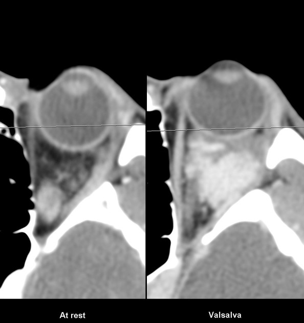
vascular
cavernous hemangioma (a.k.a. orbital venous malformation)
orbital hematoma/retrobulbar hemorrhage
-
developmental anomalies
-
paranasal sinus enlargement
-
orbital wall osseous lesions
fibro-osseous lesions: e.g fibrous dysplasia
orbital ossifying fibroma
Radiographic features
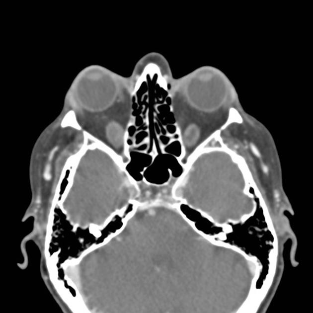
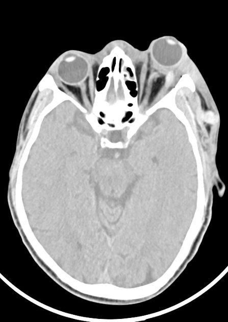
CT
Assessment of proptosis on cross-sectional imaging is difficult and dependant on the study being acquired in the correct plane:
the plane of the study must be parallel to the head of the optic nerve and the lens
the patient must have their eyes open and be looking forward with no eye movement
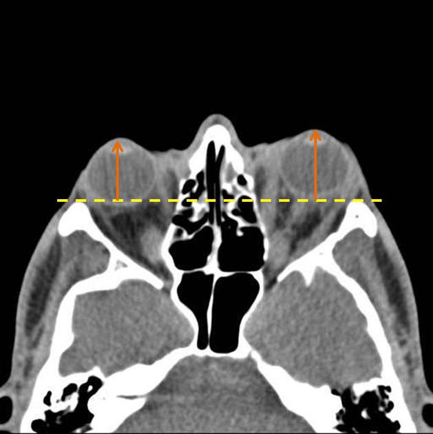
The reference line for measurement of proptosis is the interzygomatic line (a line is drawn at the anterior portions of the zygomatic bones):
the upper limit of normal distance from this line to the anterior surface of the globe is 23 mm, above which indicates proptosis 4
the lower limit of normal distance from this line to the posterior surface of the globe is 5.9 mm, below which indicates proptosis 2
The thickness of the extraocular muscles can also be used 1.
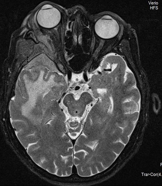
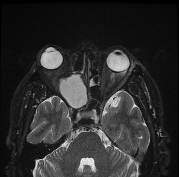
MRI
MRI may also be used in evaluation due to its multiplanar and inherent contrast capabilities. Use of MRI prevents ionizing radiation of the orbits and risk of radiation-induced cataracts. The imaging findings are similar to those described above for CT.


 Unable to process the form. Check for errors and try again.