
Right hepatic artery
Citation, DOI, disclosures and article data
At the time the article was created Stephanie BM Tan had no recorded disclosures.
View Stephanie BM Tan's current disclosuresAt the time the article was last revised Raymond Chieng had no recorded disclosures.
View Raymond Chieng's current disclosuresThe right hepatic artery (RHA) is formed when the proper hepatic artery (PHA) bifurcates. The hepatic arteries provide 25% of the blood supply and 50% of the oxygen supply to the liver.
On this page:
Images:
Gross anatomy
The proper hepatic artery bifurcates into the right and left hepatic arteries at or before reaching the porta hepatis. These are end arteries and supply the right and left halves of the liver respectively. The right hepatic artery passes upwards and turns to the right, crossing behind the common hepatic duct to enter Calot triangle 1. It usually gives off the cystic artery within Calot triangle 1,2 then turns upwards to enter the right lobe of the liver.
Within the liver, it divides into:
- anterior segmental branch which supplies segments 5 and 8 and usually supplies a branch to segment 1 1
- posterior segmental branch which supplies segments 6 and 7 1
Moynihan’s hump, also known as a caterpillar hump, is the descriptive term when the right hepatic artery forms a sinuous tortuosity occupying the majority of Calot triangle and may be at risk of damage during a cholecystectomy. It often lies in close proximity with the gallbladder neck.
NB: the hepatic segments were originally numbered by Roman numerals I to VIII, but the Arabic numerals 1 to 8 are now preferred 3
Variant anatomy
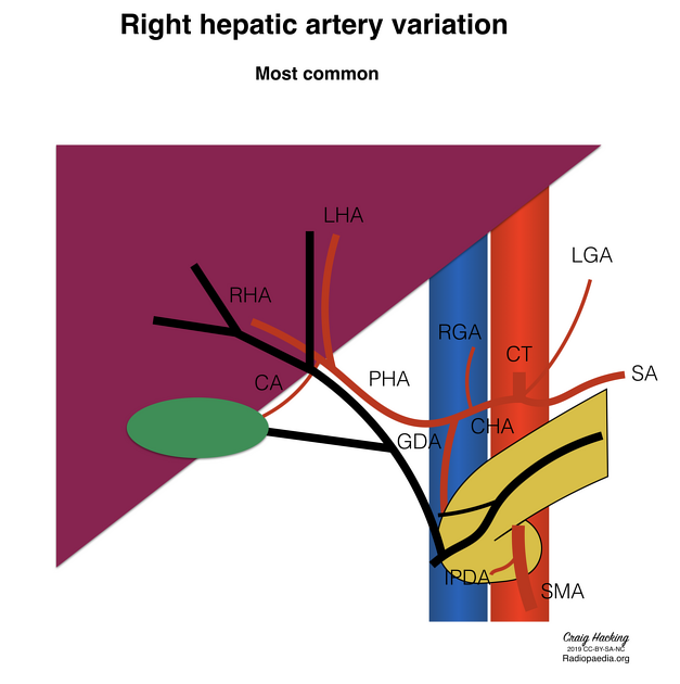
The right hepatic artery may be aberrant in one-third of cases, and is known to originate from the:
The right hepatic artery may also give rise to the middle hepatic artery, which usually arises from the left hepatic artery and supplies segments 4a and 4b.
An uncommon but important variant, the falciform artery, may arise from the right hepatic artery exiting the liver in the falciform ligament to supply part of the anterior abdominal wall.
See also
References
- 1. Dandekar U, Dandekar K, Chavan S. Right Hepatic Artery: A Cadaver Investigation and Its Clinical Significance. Anat Res Int. 16;2015: 412595. doi:10.1155/2015/412595 - Free text at pubmed - Pubmed citation
- 2. Nagral S. Anatomy relevant to cholecystectomy. J Minim Access Surg. 2005;1 (2): 53-8. doi:10.4103/0972-9941.16527 - Free text at pubmed - Pubmed citation
- 3. Strasberg SM. Nomenclature of hepatic anatomy and resections: a review of the Brisbane 2000 system. (2005) Journal of hepato-biliary-pancreatic surgery. 12 (5): 351-5. doi:10.1007/s00534-005-0999-7 - Pubmed
Incoming Links
- Hepatocystic triangle
- Bile duct stricture
- Order of structures in the porta hepatis (mnemonic)
- Coeliac artery
- Right gastric artery
- Hepatic artery proper
- Variant hepatic arterial anatomy
- Medical abbreviations and acronyms (R)
- Cystic duct
- Left hepatic artery
- Gallbladder
- Common bile duct
- Choledocholithiasis
- Magnetic resonance cholangiopancreatography (MRCP)
- Cholangiocarcinoma
- Middle hepatic artery
- Common hepatic artery
- Falciform artery
- Cystic artery
- Antiphospholipid syndrome with adrenal insufficiency
- Infiltrative hepatocellular carcinoma
- Calyceal diverticulum
- Common hepatic artery originating from aorta
- Coeliac trunk compression syndrome and aberrant left hepatic artery
- Celiacomesenteric trunk
- Ruptured internal iliac artery aneurysm
- Right hepatic artery anatomic variation (diagram)
- Hepatic arteriovenous malformation
- Hepatocellular carcinoma rupture with haemoperitoneum
Related articles: Anatomy: Abdominopelvic
- skeleton of the abdomen and pelvis
- muscles of the abdomen and pelvis
- spaces of the abdomen and pelvis
- anterior abdominal wall
- posterior abdominal wall
- abdominal cavity
- pelvic cavity
- perineum
- abdominal and pelvic viscera
- gastrointestinal tract
- spleen
- hepatobiliary system
-
endocrine system
-
adrenal gland
- adrenal vessels
- chromaffin cells
- variants
- pancreas
- organs of Zuckerkandl
-
adrenal gland
-
urinary system
-
kidney
- renal pelvis
- renal sinus
- avascular plane of Brodel
-
variants
- number
- fusion
- location
- shape
- ureter
- urinary bladder
- urethra
- embryology
-
kidney
- male reproductive system
-
female reproductive system
- vulva
- vagina
- uterus
- adnexa
- Fallopian tubes
- ovaries
- broad ligament (mnemonic)
- variant anatomy
- embryology
- blood supply of the abdomen and pelvis
- arteries
-
abdominal aorta
- inferior phrenic artery
- coeliac artery
- superior mesenteric artery
- middle suprarenal artery
- renal artery (variant anatomy)
- gonadal artery (ovarian artery | testicular artery)
- inferior mesenteric artery
- lumbar arteries
- median sacral artery
-
common iliac artery
- external iliac artery
-
internal iliac artery (mnemonic)
- anterior division
- umbilical artery
- superior vesical artery
- obturator artery
- vaginal artery
- inferior vesical artery
- uterine artery
- middle rectal artery
-
internal pudendal artery
- inferior rectal artery
-
perineal artery
- posterior scrotal artery
- transverse perineal artery
- artery to the bulb
- deep artery of the penis/clitoris
- dorsal artery of the penis/clitoris
- inferior gluteal artery
- posterior division (mnemonic)
- variant anatomy
- anterior division
-
abdominal aorta
- portal venous system
- veins
- anastomoses
- arterioarterial anastomoses
- portal-systemic venous collateral pathways
- watershed areas
- arteries
- lymphatics
- innervation of the abdomen and pelvis
- thoracic splanchnic nerves
- lumbar plexus
-
sacral plexus
- lumbosacral trunk
- sciatic nerve
- superior gluteal nerve
- inferior gluteal nerve
- nerve to piriformis
- perforating cutaneous nerve
- posterior femoral cutaneous nerve
- parasympathetic pelvic splanchnic nerves
- pudendal nerve
- nerve to quadratus femoris and inferior gemellus muscles
- nerve to internal obturator and superior gemellus muscles
- autonomic ganglia and plexuses


 Unable to process the form. Check for errors and try again.