The sacrum is the penultimate segment of the vertebral column and also forms the posterior part of the bony pelvis. It transmits the total body weight between the lower appendicular skeleton and the axial skeleton.
On this page:
Gross anatomy
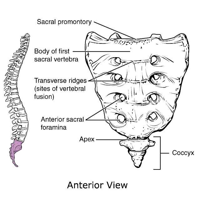
The sacrum is an irregularly shaped bone, shaped roughly like an inverted triangle, with its base superior and apex inferior. It is curved with an anterior concavity and posterior convexity.
The sacrum consists of five fused sacral vertebral and costal segments (S1-S5) that form a central sacral body and paired sacral alae (singular ala), which arise laterally from S1.
As the sacrum develops, costal elements form the parts superior, lateral and inferior to the anterior sacral foramina. Posterior sacral foramina are wholly formed by the vertebral part, the costal elements lying lateral to the lateral sacral crest.
Superiorly, the anterior lip of the S1 is called the sacral promontory. Anteriorly, the lines of vertebral fusion can be seen as four transverse ridges. Posteriorly, the spinous processes are fused to form the median sacral crest.
Through the center of the sacral body passes the triangular-shaped sacral canal, which is the continuation of the lumbar vertebral canal. It terminates inferiorly at the sacral hiatus and contains sacral and coccygeal nerve roots, spinal meninges (to the level of S2) and filum terminale. The spinal epidural space extends to the sacral hiatus. Anterior rami of the first to fourth sacral nerve roots exit via four paired anterior sacral foramina, while the smaller posterior rami exit via the four paired posterior foramina. The fifth sacral nerve root exits via the sacral hiatus.
Movements and ranges of motion at the sacrum and sacroiliac joint is limited to a few degrees maximum. Nutation and counternutation (anterior and posterior sacral tilting) are clinically important motions that occur at the sacrum, as they are capable of changing the size of the pelvic inlet and outlet during pregnancy.
Articulations
-
lumbosacral: superiorly, the sacrum articulates with the inferior endplate of L5 via the L5/S1 intervertebral disc; superior articular processes of S1 articulate with the L5 inferior articular facets to form the L5/S1 facet (zygapophyseal) joints
joint surfaces face anteroposteriorly (note interlumbar segments face laterally)
sacrococcygeal: inferiorly, the sacrum articulates with the first coccygeal segment
sacroiliac: laterally, the sacral ala articulates with the ilium
Attachments
Ligamentous
lumbosacral and iliolumbar ligaments: reinforce the lumbosacral articulation
ligamentum flavum (S1 to L5): closes the spinal/sacral canal posteriorly
sacroiliac ligaments: reinforce the sacroiliac joint - anterior, posterior and interosseous
sacrococcygeal ligaments: anterior, lateral and posterior ligaments. Superficial part of the posterior sacrococcygeal ligament closes the sacral hiatus. The anterior sacrococcygeal ligament is the terminal continuation of the anterior longitudinal ligament which blends into the periosteum of the coccyx
Musculotendinous
iliacus muscle: anterosuperiorly
piriformis muscle: anteroinferiorly
coccygeus: anteroinferiorly
erector spinae muscle: posteriorly
gluteus maximus: posteriorly
Relations
Anteriorly:
rectosacral fascia of Waldeyer, presacral fascia, peritoneum (S1-2), rectum (S3-5), lymph nodes
-
in the midline from above down:
median sacral vessels
superior rectal artery
-
on each side of the midline
sympathetic chain
lateral sacral vessels
Arterial supply
iliolumbar arteries (posterior division of internal iliac artery)
superior and inferior lateral sacral arteries (posterior division of internal iliac artery)
Venous drainage
-
via vertebral venous plexus to:
median sacral vein (to either common iliac vein or IVC)
lateral sacral vein (to internal iliac vein)
Variant anatomy
Read more in our article on anatomical variants of the sacrum