
Sheehan syndrome is a rare cause of pituitary apoplexy and hypopituitarism. It only occurs in postpartum females who experience large volume haemorrhage and hypovolaemic shock, either during delivery or afterward with resultant necrosis of anterior pituitary cells 4.
On this page:
Epidemiology
Advances in obstetrical care mean Sheehan syndrome is rare in developed countries. The incidence in developing and low-income countries is as high as 5 in 100,000 births 5.
Clinical presentation
-
pituitary failure 1
may be silent and present with delayed hypopituitarism
agalactorrhoea
amenorrhoea
oligomenorrhoea
adrenal insufficiency
hypernatraemia (diabetes insipidus) in the acute setting of extreme hypovolaemia
hypothyroidism
growth hormone deficiency
-
optic chiasm compression
visual field loss
headache
Pathology
Hyperplasia of pituitary cells, particularly lactotrophs, occurs in the weeks preceding delivery resulting in an increased metabolic demand without a concomitant increase in blood supply 4. The anterior pituitary blood supply is under relatively low pressure making these cells susceptible to ischaemia. Pregnancies complicated by hypovolaemia secondary to postpartum haemorrhage may lead to pituitary infarction and necrosis 4.
The posterior pituitary has its blood supply under higher pressure and is not usually affected however diabetes insipidus may occur as a rare manifestation of Sheehan syndrome 4,5.
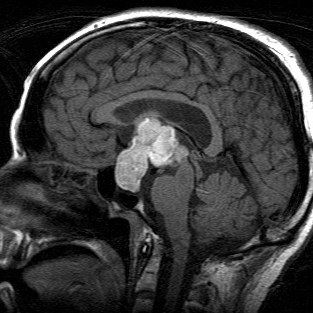
Radiographic features
CT
ring enhancement surrounding a low attenuation empty sella 2
MRI
-
early
enlarged pituitary, with low T1, high T2 homogenous signal
ring enhancement
-
late
empty sella of normal size 3
Treatment and prognosis
The basis of treatment is lifelong replacement of deficient hormones. Anterior pituitary hormones are generally affected in the following order after necrosis; growth hormone, prolactin, follicle-stimulating hormone, luteinizing hormone, adrenocorticotropic hormone and then thyroid-stimulating hormone 5.
History and etymology
It is named after Harold Leeming Sheehan (1900-1988), a British pathologist who first described the syndrome in 1937 5.


 Unable to process the form. Check for errors and try again.