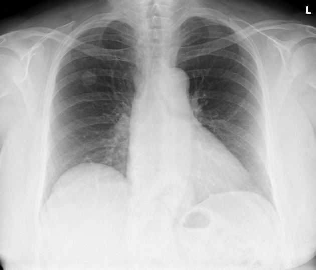
A solitary pulmonary nodule, according to the Nomenclature Committee of the Fleischner Society, is defined as a rounded opacity, well or poorly-defined on a conventional radiograph, measuring up to 3 cm in diameter and is not associated with lymphadenopathy, atelectasis, or pneumonia.
Several radiographic parameters have been described for the risk assessment of a lung nodule to include or exclude possible chances of malignancy. These radiographic features must be kept in the mind while evaluating a solitary pulmonary nodule:
Nodule size
Generally, a nodule greater than 7 mm in a patient above 40 years of age is considered to be associated with increased risk of malignancy. Nodule size and risk of malignancy observed:
- <5 mm: <1%
- 5-10 mm: 6-28%
- >20 mm: 64-82%
Nodule growth rate
If previous radiographs are available, the growth rate of a nodule can be calculated in terms of doubling time. Cancerous lesions have a doubling time of 1-18 months.
Nodule location
Benign lesions like granulomas and intrapulmonary lymph nodes generally occur more peripherally, particularly in subpleural regions. Most of the primary lung cancer affect upper lobe (right>left) and majority of metastatic lesions show lower lobe predominance.
Nodule margins
Well-defined and smooth margins are features of benign lesions while in malignant lesions, margins are more ill-defined/spiculated/undulated due to desmoplastic reaction or infiltration of interstitium by the tumour cells.
Calcification within nodule
Various types of calcifications within the nodule have been described. Dense, solid and laminated calcifications are most commonly seen in old granulomas. Popcorn calcification is a feature of hamartoma. Calcifications in malignant nodules are fine, dendriform and punctate.
Fat within nodule
The presence of fat within a nodule is a strong indicator of benignity and seen with hamartoma, lipoma and lipoid pneumonia.
Cavitation within nodule
Cavitation within nodule increases the risk of possible malignancy especially if the cavity is thick-walled and irregular in shape. Although benign lesions may also cavitate but these cavities are generally thin-walled and round.
Attenuation
Partly solid nodules are more likely to be malignant than pure solid or fluid-containing nodules.
CT densitometry
High-density lesions on CT are more likely to be benign.
Enhancement
Enhancement is related to vascularity and more enhancement is associated with increased risk. Malignant lesions show average enhancement of 40 HU (20-108 HU) which is significantly higher than that of benign granulomas of average 12 HU (-4-58 HU). An enhancement less than 15 HU is highly suggestive of benignity.
Dynamic contrast-enhanced MRI has shown that malignant nodules show early peak enhancement and faster washout.
Feeding vessel sign
The feeding vessel sign is frequently associated with metastatic nodules, however, it can also be seen commonly with septic emboli.


 Unable to process the form. Check for errors and try again.