Subcutaneous emphysema
Updates to Article Attributes
Subcutaneous or subcutaneous emphysema, strictly speaking, refers to air in the subcutaneous tissues. But the term is generally used to describe any soft tissue emphysema of the body wall or limbs, since the air often dissects into the deeper soft tissues and musculature along fascial planes.
Clinical presentation
Clinically it is felt as crepitus and, if extensive, may cause soft tissue swelling and discomfort. Even when severe subcutaneous emphysema is typically benign, although complications such as airway compromise, respiratory failure, pacemaker malfunction and tension phenomena have been described.
In the trauma situation, the gas often does not need treatment itself, but its importance lies in the fact that its presence indicates possible serious injuries that does require urgent management. Air can track along fascial planes and enter the head, neck, liumbs, chest, abdomen, and scrotum.
Pathology
Causes of subcutaneous emphysema can be divided into:
- gas arising internally
- pneumothorax
- pneumomediastinum
- pulmonary interstitial emphysema
- perforated hollow viscus in the neck, e.g. oesophageal perforation
- fistula tract
- gas introduced externally
- penetrating trauma
- post-surgical
- post-percutaneous intervention, e.g. pleural drain insertion
-
gas produced de novo
- gas producing infection(s), e.g necrotising fasciitis
Trauma is the most common cause seen 5.
Radiographic appearance
Plain film
There is often striated lucencies in the soft tissues that may outline muscle fibres. If affecting the anterior chest wall, subcutaneous emphysema can outline the pectoralis major muscle, giving rise to the ginkgo leaf sign 2. Often there is displaced rib fractures indicating a cause of the gas.
CT chest
Subcutaneous emphysema is readily visible on CT scans, with pockets of air seen as extremely dark low (air) attenuation areas in the subcutaneous space.
Treatment and prognosis
Treatment is directed at the underlying cause, while the subcutaneous air is absorbed by the body over time. Symptomatic management should also be provided.
However in rare instances where the subcutaneous air is compromising overlying soft tissue or causing a compartment syndrome management may involve release of the air by surgical division of the soft tissues or percutaneous drain insertion.
Differential diagnosis
- gas trapped in skin folds or clothing
- gas within soft tissue lacerations
- gas associated with long hair
-</ul><p>Trauma is the most common cause seen <sup>5</sup>.</p><h4>Radiographic appearance</h4><h5>Plain film</h5><p>There is often striated lucencies in the soft tissues that may outline muscle fibres. If affecting the anterior chest wall, subcutaneous emphysema can outline the <a href="/articles/pectoralis-major-1">pectoralis major muscle</a>, giving rise to the <a href="/articles/ginkgo-leaf-sign-chest-x-ray">ginkgo leaf sign</a> <sup>2</sup>. Often there is displaced rib fractures indicating a cause of the gas.</p><h5>CT chest</h5><p>Subcutaneous emphysema is readily visible on CT scans, with pockets of air seen as extremely dark low (air) attenuation areas in the subcutaneous space.</p><h4>Treatment and prognosis</h4><p>Treatment is directed at the underlying cause, while the subcutaneous air is absorbed by the body over time. Symptomatic management should also be provided.</p><p>However in rare instances where the subcutaneous air is compromising overlying soft tissue or causing a compartment syndrome management may involve release of the air by surgical division of the soft tissues or percutaneous drain insertion.</p><h4>Differential diagnosis</h4><ul>- +</ul><p>Trauma is the most common cause seen <sup>5</sup>.</p><h4>Radiographic appearance</h4><h5>Plain film</h5><p>There is often striated lucencies in the soft tissues that may outline muscle fibres. If affecting the anterior chest wall, subcutaneous emphysema can outline the <a href="/articles/pectoralis-major-1">pectoralis major muscle</a>, giving rise to the <a href="/articles/ginkgo-leaf-sign-chest">ginkgo leaf sign</a> <sup>2</sup>. Often there is displaced rib fractures indicating a cause of the gas.</p><h5>CT chest</h5><p>Subcutaneous emphysema is readily visible on CT scans, with pockets of air seen as extremely dark low (air) attenuation areas in the subcutaneous space.</p><h4>Treatment and prognosis</h4><p>Treatment is directed at the underlying cause, while the subcutaneous air is absorbed by the body over time. Symptomatic management should also be provided.</p><p>However in rare instances where the subcutaneous air is compromising overlying soft tissue or causing a compartment syndrome management may involve release of the air by surgical division of the soft tissues or percutaneous drain insertion.</p><h4>Differential diagnosis</h4><ul>
Image 1 CT (Scout) ( create )
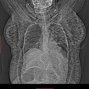
Image 2 CT (lung window) ( update )
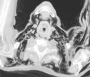
Image 3 X-ray (Frontal) ( update )

Image 4 X-ray ( update )
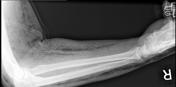
Image 5 X-ray (Frontal) ( update )

Image 6 CT (C+ portal venous phase) ( update )

Image 7 X-ray (Frontal) ( update )

Image 8 CT (lung window) ( update )

Image 9 X-ray (Frontal, supine AP) ( update )

Image 10 CT (lung window) ( update )

Image 11 CT (lung window) ( update )

Image 12 CT (lung window) ( update )
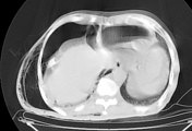
Image 13 X-ray (Supine AP) ( update )

Image 14 X-ray (Frontal) ( update )
