
Tailgut cysts, also known as retrorectal cystic hamartomas, are rare congenital lesions that are thought to arise from vestiges of the embryonic hindgut.
On this page:
Epidemiology
There is a recognized strong female predilection. While it can be presented at any age, presentation is usually at around 30-60 years of age 4.
Clinical presentation
Many lesions are discovered incidentally 5. Approximately 50% of patients may have perirectal symptoms, likely pelvic pain and constipation 9,10.
Pathology
On gross pathological examination, a tailgut cyst usually consists of a multiloculated, cystic mass with a thin wall, glistening lining and filled with mucoid material. The cysts can be lined by a variety of epithelial cells, including ciliated columnar, mucin-secreting columnar, transitional, and squamous epithelium 10.
The lesions usually measure several centimeters in diameter. Occasionally, a sacral bone defect and/or associated calcifications may be present 3.
Location
It is almost exclusively found in the retrorectal or presacral space and very rarely in other sites such as the perirenal area or the subcutaneous tissues 7.
Radiographic features
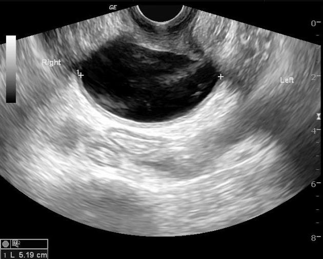
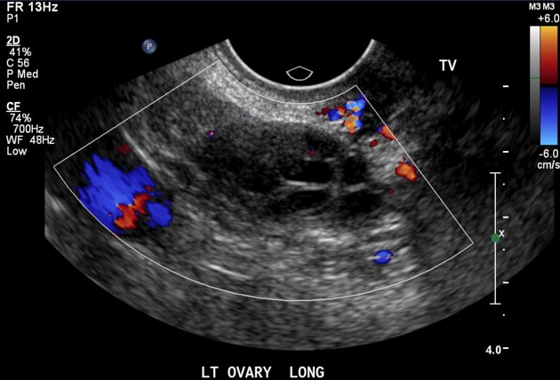
Ultrasound
Transrectal ultrasound may show a multilocular, retrorectal cystic mass. Internal echoes may be found within the cyst due to the multi-cystic nature of the mass and the presence of gelatinous material or inflammatory debris within the cyst.
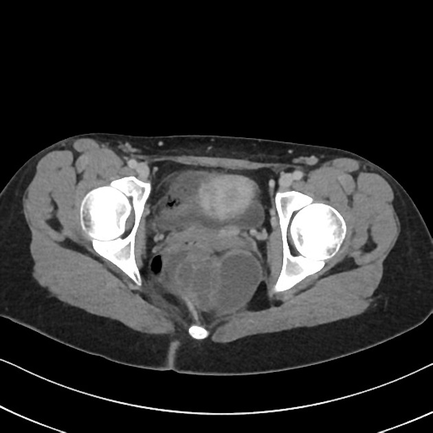
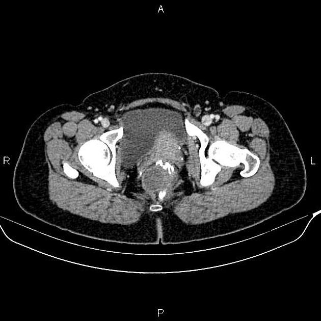
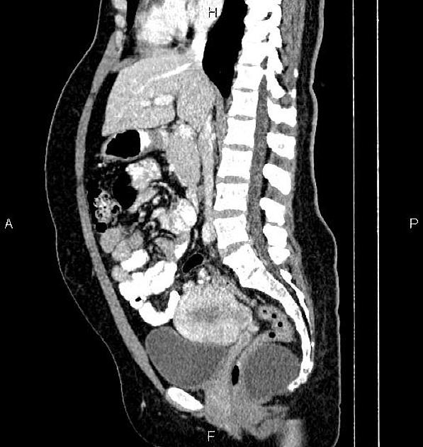
CT
Often seen as a discrete, well-marginated, presacral mass with water or soft-tissue density, depending on the contents of the cyst. Calcifications may be seen in the cyst wall. When the mass is large, the rectum is displaced by the mass. If concurrent infection or malignant transformation occurs, CT may reveal loss of discrete margins and involvement of contiguous structures.
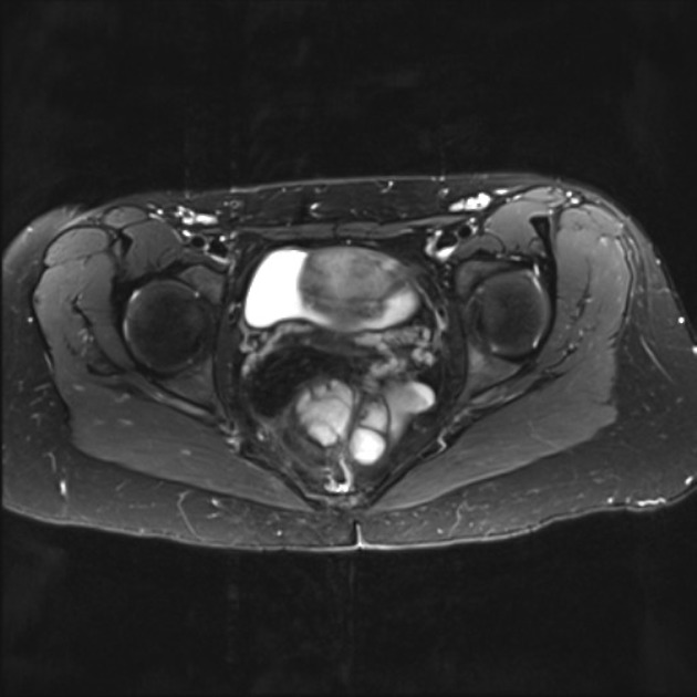
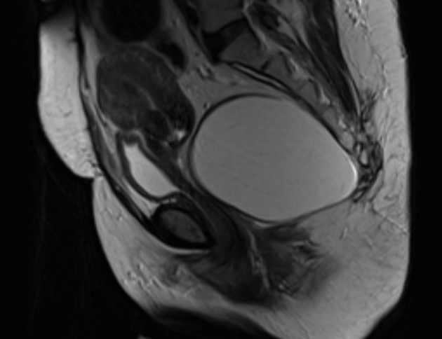
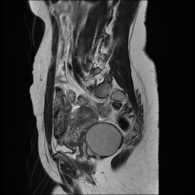
MRI
MRI signal characteristics depend on whether the cyst is complicated or not. Complications include:
infection or inflammation
hemorrhage 14
malignant change: rare and concerning potential complication 1
Uncomplicated cysts
T1: low signal
-
T2: high signal
some reports suggest that a multilocular appearance with internal septa on T2 images to a cyst in the retrorectal region is a feature unique to tailgut cysts 6
Complicated cysts
T1: high signal components may occur due to the presence of mucinous material, high protein content, or associated intracystic hemorrhage
T2: low signal components may occur due to the presence of hemorrhage or associated keratin 4
Treatment and prognosis
While uncomplicated cysts are benign, surgical excision is the recommended treatment of choice even in asymptomatic cases, especially because of potential complications 3-5.
Differential diagnosis
General imaging differential considerations for a cystic lesion in the retrorectal region are rather broad and include 3,8:
-
other developmental cysts in the retrorectal region
cystic sacrococcygeal teratoma
anal duct cyst: anal gland cyst
cystic lymphangioma in the retrorectal region
retrorectal pyogenic abscess
necrotic sacral chordoma


 Unable to process the form. Check for errors and try again.