
Thermoluminescent dosimeter
Citation, DOI, disclosures and article data
At the time the article was created Ayush Goel had no recorded disclosures.
View Ayush Goel's current disclosuresAt the time the article was last revised Mateusz Wilczek had no financial relationships to ineligible companies to disclose.
View Mateusz Wilczek's current disclosures- TLD
- Thermoluminescent dosimeter badge
- TLD badge
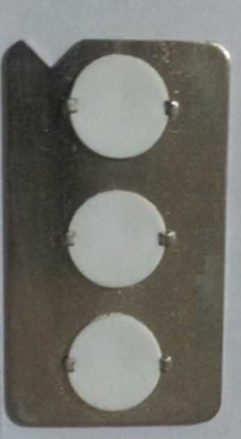
Thermoluminescent dosimeter (TLD) is a passive radiation detection device that is used for personal dose monitoring or to measure patient dose.
Parts
plastic holder
-
nickel-coated aluminium card with TLD discs
-
the discs are made of a thermoluminescent material, commonly calcium sulphate doped with dysprosium (CaSO4:Dy) or lithium fluoride (LiF)
nearly tissue equivalent, although not at all x-ray energies 1
the discs are 0.8 mm thick and have a 1.35 cm diameter
-
-
three filters against each disc
top: aluminium and copper
middle: perspex
lower: open
When the radiation falls on TLD, the electrons are excited and store energy. After a varied period (can be one month, bimonthly, quarterly or even biannual), the TLD badges are sent for reading. The TLD reader consists of a heater. On getting heated, the excited electrons again come back to the ground state and emit light which is then read by a photomultiplier 2. The light output is proportional to the radiation exposure 3.
TLD has better accuracy than a film dosimeter and do not require an estimate of photon energy. It also has a higher dynamic range and are re-usable 3.
If a radiation worker is wearing a lead apron, the TLD badge can be worn under the lead apron (to reflect body dose) or outside the lead apron (to reflect extremity/thyroid dose) 2. TLDs can measure doses from 0.01 mGy to 10 Gy 3.
Quiz questions
References
- 1. Jamie V. Trapp, Tomas Kron. An Introduction to Radiation Protection in Medicine. ISBN: 9781584889656
- 2. Curry TS, Dowdey JE, Murry RE. Christensen ́s physics of diagnostic radiology 4 Ed. Lippincott Williams & Wilkins. (1990) ISBN:0812113101. Read it at Google Books - Find it at Amazon
- 3. Walter Huda. Review of Radiologic Physics. ISBN: 9780781785693
Incoming Links
Related articles: Imaging technology
- imaging technology
- imaging physics
- imaging in practice
-
x-rays
- x-ray physics
- x-ray in practice
- x-ray production
- x-ray tube
- filters
- automatic exposure control (AEC)
- beam collimators
- grids
- air gap technique
- cassette
- intensifying screen
- x-ray film
- image intensifier
- digital radiography
- digital image
- mammography
- x-ray artifacts
- radiation units
- radiation safety
- radiation detectors
- fluoroscopy
-
computed tomography (CT)
- CT physics
- CT in practice
- CT technology
- CT image reconstruction
- CT image quality
- CT dose
-
CT contrast media
-
iodinated contrast media
- agents
- water soluble
- water insoluble
- vicarious contrast material excretion
- iodinated contrast media adverse reactions
- agents
- non-iodinated contrast media
-
iodinated contrast media
-
CT artifacts
- patient-based artifacts
- physics-based artifacts
- hardware-based artifacts
- ring artifact
- tube arcing
- out of field artifact
- air bubble artifact
- helical and multichannel artifacts
- CT safety
- history of CT
-
MRI
- MRI physics
- MRI in practice
- MRI hardware
- signal processing
-
MRI pulse sequences (basics | abbreviations | parameters)
- T1 weighted image
- T2 weighted image
- proton density weighted image
- chemical exchange saturation transfer
- CSF flow studies
- diffusion weighted imaging (DWI)
- echo-planar pulse sequences
- fat-suppressed imaging sequences
- gradient echo sequences
- inversion recovery sequences
- metal artifact reduction sequence (MARS)
-
perfusion-weighted imaging
- techniques
- derived values
- saturation recovery sequences
- spin echo sequences
- spiral pulse sequences
- susceptibility-weighted imaging (SWI)
- T1 rho
- MR angiography (and venography)
-
MR spectroscopy (MRS)
- 2-hydroxyglutarate peak: resonates at 2.25 ppm
- alanine peak: resonates at 1.48 ppm
- choline peak: resonates at 3.2 ppm
- citrate peak: resonates at 2.6 ppm
- creatine peak: resonates at 3.0 ppm
- functional MRI (fMRI)
- gamma-aminobutyric acid (GABA) peak: resonates at 2.2-2.4 ppm
- glutamine-glutamate peak: resonates at 2.2-2.4 ppm
- Hunter's angle
- lactate peak: resonates at 1.3 ppm
- lipids peak: resonates at 1.3 ppm
- myoinositol peak: resonates at 3.5 ppm
- MR fingerprinting
- N-acetylaspartate (NAA) peak: resonates at 2.0 ppm
- propylene glycol peak: resonates at 1.13 ppm
-
MRI artifacts
- MRI hardware and room shielding
- MRI software
- patient and physiologic motion
- tissue heterogeneity and foreign bodies
- Fourier transform and Nyquist sampling theorem
- MRI contrast agents
- MRI safety
-
ultrasound
- ultrasound physics
-
transducers
- linear array
- convex array
- phased array
- frame averaging (frame persistence)
- ultrasound image resolution
- imaging modes and display
- pulse-echo imaging
- real-time imaging
-
Doppler imaging
- Doppler effect
- colour Doppler
- power Doppler
- B flow
- colour box
- Doppler angle
- pulse repetition frequency and scale
- wall filter
- colour write priority
- packet size (dwell time)
- peak systolic velocity
- end-diastolic velocity
- resistive index
- pulsatility index
- Reynolds number
- panoramic imaging
- compound imaging
- harmonic imaging
- elastography
- scanning modes
- 2D ultrasound
- 3D ultrasound
- 4D ultrasound
- M-mode
-
ultrasound artifacts
- acoustic shadowing
- acoustic enhancement
- beam width artifact
- reverberation artifact
- ring down artifact
- mirror image artifact
- side lobe artifact
- speckle artifact
- speed displacement artifact
- refraction artifact
- multipath artifact
- anisotropy
- electrical interference artifact
- hardware-related artifacts
- Doppler artifacts
- aliasing
- tissue vibration
- spectral broadening
- blooming
- motion (flash) artifact
- twinkling artifact
- acoustic streaming
- biological effects of ultrasound
- history of ultrasound
-
nuclear medicine
- nuclear medicine physics
- detectors
- tissue to background ratio
-
radiopharmaceuticals
- fundamentals of radiopharmaceuticals
- radiopharmaceutical labelling
- radiopharmaceutical production
- nuclear reactor produced radionuclides
- cyclotron produced radionuclides
- radiation detection
- dosimetry
- specific agents
- carbon-11
- chromium-51
- fluorine agents
- gallium agents
- Ga-67 citrate
- Ga-68
- iodine agents
-
I-123
- I-123 iodide
- I-123 ioflupane (DaTSCAN)
- I-123 ortho-iodohippurate
- I-131
-
MIBG scans
- I-123 MIBG
- I-131 MIBG
-
I-123
- indium agents
- In-111 Octreoscan
- In-111 OncoScint
- In-111 Prostascint
- In-111 oxine labelled WBC
- krypton-81m
- nitrogen-13
- oxygen-15
- phosphorus-32
- selenium-75
-
technetium agents
- Tc-99m DMSA
- Tc-99m DTPA
- Tc-99m DTPA aerosol
- Tc-99m HMPAO
- Tc-99m HMPAO labelled WBC
- Tc-99m MAA
- Tc-99m MAG3
- Tc-99m MDP
- Tc-99m mercaptoacetyltriglycine
- Tc-99m pertechnetate
- Tc-99m labelled RBC
- Tc-99m sestamibi
- Tc-99m sulfur colloid
- Tc-99m sulfur colloid (oral)
- thallium-201 chloride
- xenon agents
- in vivo therapeutic agents
- pharmaceuticals used in nuclear medicine
-
emerging methods in medical imaging
- radiography
- phase-contrast imaging
- CT
- deep-learning reconstruction
- photon counting CT
- virtual non-contrast imaging
- ultrasound
- magnetomotive ultrasound (MMUS)
- superb microvascular imaging
- ultrafast Doppler imaging
- ultrasound localisation microscopy
- MRI
- nuclear medicine
- total body PET system
- immuno-PET
- miscellaneous
- radiography


 Unable to process the form. Check for errors and try again.