
Thyroglossal duct
Citation, DOI, disclosures and article data
At the time the article was created Frank Gaillard had no recorded disclosures.
View Frank Gaillard's current disclosuresAt the time the article was last revised Calum Worsley had no financial relationships to ineligible companies to disclose.
View Calum Worsley's current disclosures- TD
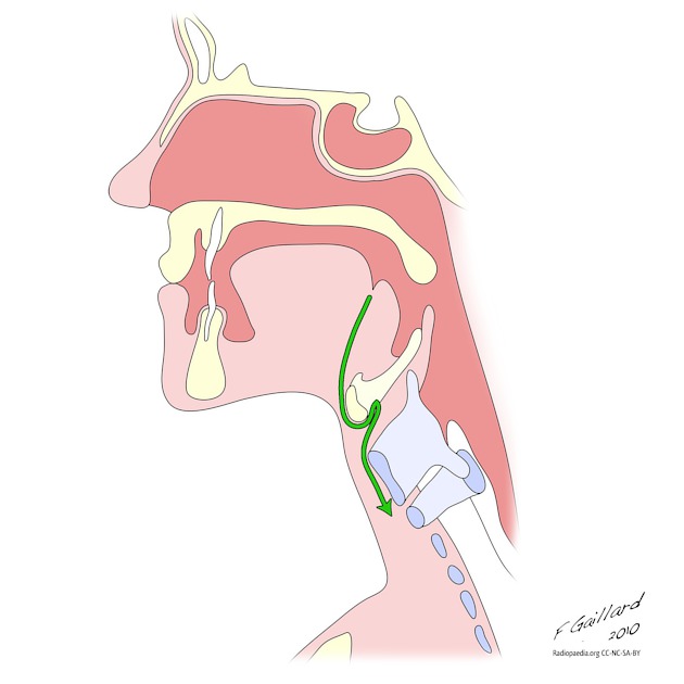
The thyroglossal duct is an epithelium-lined connection between the foramen cecum and the thyroid that forms during the descent of the thyroid during embryological development. It usually involutes in the 8th-10th week of gestation.
On this page:
Gross anatomy
The thyroglossal duct arises from proximal primitive foregut between the first and second pharyngeal pouches at the foramen cecum, located at the junction of the anterior two thirds and posterior third of the tongue 1. From there it passes anterior to the body of the hyoid bone, curving backwards and superiorly to reach behind the bone before once more turning inferiorly and continuing to the isthmus of the thyroid. The pyramidal lobe, if present, marks this point. The tip of the duct bifurcates, forming the two lobes of the gland. The parafollicular cells (C cells) responsible for calcitonin production are derived from separate tissue, the ultimobranchial body, a small diverticulum of the fourth pharyngeal pouch.
Variant anatomy
The thyroid may halt at any stage during its descent or leave rests of thyroid tissue along the course of the thyroglossal duct 2:
Related pathology
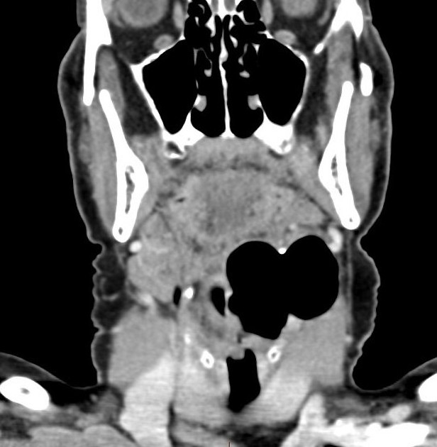
Failure of obliteration of the duct may lead to the formation of a thyroglossal duct cyst.
References
- 1. Foley D & Fallat M. Thyroglossal Duct and Other Congenital Midline Cervical Anomalies. Semin Pediatr Surg. 2006;15(2):70-5. doi:10.1053/j.sempedsurg.2006.02.003 - Pubmed
- 2. Ranade A, Rai R, Pai M et al. Anatomical Variations of the Thyroid Gland: Possible Surgical Implications. Singapore Med J. 2008;49(10):831-4. - Pubmed
- 3. Branstetter B, Weissman J, Kennedy T, Whitaker M. The CT Appearance of Thyroglossal Duct Carcinoma. AJNR Am J Neuroradiol. 2000;21(8):1547-50. PMC7974032 - Pubmed
- 4. Last, R. J., McMinn, R. M. H.. Last's Anatomy, Regional and Applied. (1994) ISBN: 044304662X - Google Books
- 5. Susan Standring. Gray's Anatomy. (2015) ISBN: 9780702052309 - Google Books
Incoming Links
- Thyroglossal duct cyst
- Thyroglossal duct cyst
- Hypertrophied residual thyroid tissue along thyroglossal tract following previous thyroidectomy
- Thyroglossal duct fistula
- Thyroglossal duct cyst
- Thyroglossal duct cyst
- Thyroglossal duct cyst
- Thyroglossal duct cyst with pre-epiglottic space extension
- Thyroglossal duct sinus
- Thyroglossal duct cyst
- Thyroglossal duct sinus
- Thyroglossal duct cyst
- Thyroglossal duct cyst
- Marine-Lenhart syndrome
- Thyroglossal duct cyst
- Thyroglossal duct (diagram)
- Congenital hypothyroidism - lingual thyroid
Related articles: Anatomy: Head and neck
- skeleton of the head and neck
-
cranial vault
- scalp (mnemonic)
- fontanelle
-
sutures
- calvarial
- facial
- frontozygomatic suture
- frontomaxillary suture
- frontolacrimal suture
- frontonasal suture
- temporozygomatic suture
- zygomaticomaxillary suture
- parietotemporal suture (parietomastoid suture)
- occipitotemporal suture (occipitomastoid suture)
- sphenofrontal suture
- sphenozygomatic suture
- spheno-occipital suture (not a true suture)
- lacrimomaxillary suture
- nasomaxillary suture
- internasal suture
- basal/internal
- skull landmarks
- frontal bone
- temporal bone
- parietal bone
- occipital bone
- skull base (foramina)
-
facial bones
- midline single bones
- paired bilateral bones
- cervical spine
- hyoid bone
- laryngeal cartilages
-
cranial vault
- muscles of the head and neck
- muscles of the tongue (mnemonic)
- muscles of mastication
-
facial muscles
- epicranius muscle
- circumorbital and palpebral muscles
- nasal muscles
-
buccolabial muscles
- elevators, retractors and evertors of the upper lip
- levator labii superioris alaeque nasalis muscle
- levator labii superioris muscle
- zygomaticus major muscle
- zygomaticus minor muscle
- levator anguli oris muscle
- malaris muscle
- risorius muscle
- depressors, retractors and evertors of the lower lip
- depressor labii inferioris muscle
- depressor anguli oris muscle
- mentalis muscle
- compound sphincter
-
orbicularis oris muscle
- incisivus labii superioris muscle
- incisivus labii inferioris muscle
-
orbicularis oris muscle
- muscle of mastication
- modiolus
- elevators, retractors and evertors of the upper lip
- muscles of the middle ear
- orbital muscles
- muscles of the soft palate
- pharyngeal muscles
- suprahyoid muscles
- infrahyoid muscles
- intrinsic muscles of the larynx
- muscles of the neck
- platysma muscle
- longus colli muscle
- longus capitis muscle
- scalenus anterior muscle
- scalenus medius muscle
- scalenus posterior muscle
- scalenus pleuralis muscle
- sternocleidomastoid muscle
-
suboccipital muscles
- rectus capitis posterior major muscle
- rectus capitis posterior minor muscle
- obliquus capitis superior muscle
- obliquus capitis inferior muscle
- accessory muscles of the neck
- deep cervical fascia
-
deep spaces of the neck
- anterior cervical space
- buccal space
- carotid space
- danger space
- deep cervical fascia
- infratemporal fossa
- masticator space
- parapharyngeal space
- stylomandibular tunnel
- parotid space
- pharyngeal (superficial) mucosal space
- perivertebral space
- posterior cervical space
- pterygopalatine fossa
- retropharyngeal space
- suprasternal space (of Burns)
- visceral space
- surgical triangles of the neck
- orbit
- ear
- paranasal sinuses
- upper respiratory tract
- viscera of the neck
- blood supply of the head and neck
-
arterial supply
-
common carotid artery
- carotid body
- carotid bifurcation
- subclavian artery
- variants
-
common carotid artery
- venous drainage
-
arterial supply
- innervation of the head and neck
-
cranial nerves
- olfactory nerve (CN I)
- optic nerve (CN II)
- oculomotor nerve (CN III)
- trochlear nerve (CN IV)
-
trigeminal nerve (CN V) (mnemonic)
- trigeminal ganglion
- ophthalmic division
- maxillary division
- mandibular division
- abducens nerve (CN VI)
- facial nerve (CN VII)
-
vestibulocochlear nerve (CN VIII)
- vestibular ganglion (Scarpa's ganglion)
- glossopharyngeal nerve (CN IX)
- vagus nerve (CN X)
- (spinal) accessory nerve (CN XI)
- hypoglossal nerve (CN XII)
- parasympathetic ganglia of the head and neck
- cervical sympathetic ganglia
- greater occipital nerve
- third occipital nerve
-
cervical plexus
- muscular branches
- longus capitis
- longus colli
- scalenes
- geniohyoid
- thyrohyoid
-
ansa cervicalis
- omohyoid (superior and inferior bellies separately)
- sternothyroid
- sternohyoid
- phrenic nerve
- contribution to the accessory nerve (CN XI)
- cutaneous branches
- muscular branches
- brachial plexus
- pharyngeal plexus
-
cranial nerves
- lymphatic drainage of the head and neck
- embryological development of the head and neck


 Unable to process the form. Check for errors and try again.