
Tibialis posterior muscle
Citation, DOI, disclosures and article data
At the time the article was created Jeremy Jones had no recorded disclosures.
View Jeremy Jones's current disclosuresAt the time the article was last revised Arlene Campos had no financial relationships to ineligible companies to disclose.
View Arlene Campos's current disclosures- Tibialis posterior
- Posterior tibialis tendon (PTT)
- Tibialis posterior (TP) muscle
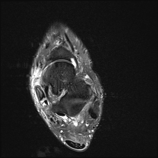
The tibialis posterior muscle is one of the small muscles of the deep posterior compartment of the leg.
Summary
origin: upper half of posterior shaft of the tibia and upper half of fibula between the medial crest and interosseous border, and adjacent interosseous membrane.
-
insertion: navicular and medial cuneiform
-
the tendon splits into two slips after passing inferior to the plantar calcaneonavicular ligament
-
superficial slip inserts on the tuberosity of the navicular bone and sometimes medial cuneiform
this is the main slip, accounting anterior two-thirds of the tendon
deeper slip divides again into slips and has variable insertions onto the plantar surfaces of metatarsals 2 - 4, intermediate cuneiform, cuboid, sustentaculum tali
-
-
action: plantarflexion and inversion of the foot
arterial supply: posterior tibial artery
innervation: tibial nerve
antagonist: tibialis anterior
-
variants:
insertion into accessory navicular
Related pathology
References
- 1. Susan Standring. Gray's Anatomy. (2008) ISBN: 9780443066849 - Google Books
- 2. Flores D, Mejía Gómez C, Fernández Hernando M, Davis M, Pathria M. Adult Acquired Flatfoot Deformity: Anatomy, Biomechanics, Staging, and Imaging Findings. Radiographics. 2019;39(5):1437-60. doi:10.1148/rg.2019190046 - Pubmed
Incoming Links
- Deep posterior compartment of the leg
- Duchenne muscular dystrophy
- Medioplantar oblique ligament
- Sciatic nerve motor distribution
- Lateral ankle sprain
- Fibular artery
- Cuboid
- Tendon instability
- Muscles of the lower limb
- Metatarsal
- Inferoplantar longitudinal ligament
- Intermediate cuneiform
- Tibia
- Ankle joint
- Tibialis posterior dysfunction
- Transverse arch of the foot
- MRI of the ankle (an approach)
- Cuboideonavicular joint
- Fibula
- Superomedial calcaneonavicular ligament
- Posterior tibialis tendon dislocation
- Partial tear of the posterior tibial tendon
- Tibialis posterior dysfunction
- Foreign body - foot
- Tibialis posterior calcific tendinosis
- Plantar ligaments of the foot (Gray's illustration)
- Spring ligament injury
- Tibialis posterior tenosynovitis
- Tibialis posterior tendon rupture
- Tibialis posterior tenosynovitis
- Navicular fracture
- Tibialis posterior tear
- Tibialis posterior tendon anatomy: diagram
- Foot muscle insertions (Gray's anatomy illustration)
- Lateral extra-articular hindfoot impingement with posterior tibial tendon split tear
- Tibialis posterior tendon rupture
- Tibialis posterior tendon tear
Related articles: Anatomy: Lower limb
- skeleton of the lower limb
- joints of the lower limb
-
hip joint
- ligaments
- muscles
- additional structures
- hip joint capsule
- zona orbicularis
- iliotibial band
-
hip bursae
- anterior
- iliopsoas bursa (iliopectineal bursa)
- lateral
- subgluteal bursae
- greater trochanteric bursa (subgluteus maximus bursa)
- subgluteus medius bursa
- subgluteus minimus bursa
- gluteofemoral bursa
- subgluteal bursae
- postero-inferior
- anterior
- ossification centres
-
knee joint
- ligaments
- anterior cruciate ligament
- posterior cruciate ligament
- medial collateral ligament
- lateral collateral ligament
- meniscofemoral ligament (mnemonic)
-
posterolateral ligamentous complex
- arcuate ligament
- patellar tendon and quadriceps tendon
- anterolateral ligament
- posterior oblique ligament
- oblique popliteal ligament
- medial patellofemoral ligament
- additional structures
- extensor mechanism of the knee
- groove for the popliteus tendon
- knee bursae
- anterior bursae
- medial bursae
- lateral bursae
- posterior bursae
- knee capsule
- lateral patellar retinaculum
- medial patellar retinaculum
- menisci
- pes anserinus (mnemonic)
- ossification centres
- ligaments
- tibiofibular joints
-
ankle joint
- regional anatomy
- medial ankle
- lateral ankle
- anterior ankle
- ligaments
- medial collateral (deltoid) ligament
- lateral collateral ligament
- additional structures
- ankle bursae
- ossification centres of the ankle
- variants
- regional anatomy
- foot joints
- subtalar joint
- mid-tarsal (Chopart) joint
-
tarsometatarsal (Lisfranc) joint
- ligaments
- intermetatarsal joint
- metatarsophalangeal joint
- interphalangeal joint
- ossification centres
-
hip joint
- spaces of the lower limb
-
muscles of the lower limb
- muscles of the pelvic group
- muscles of the thigh
- muscles of the leg
- anterior compartment of the leg
- posterior compartments of the leg
- lateral compartment of the leg
- muscles of the foot
- dorsal muscles
- plantar muscles
- 1st layer
- 2nd layer
- 3rd layer
- 4th layer
- accessory muscles of the lower limb
- accessory gluteal muscles
-
accessory muscles of the ankle
- accessory peroneal muscles
- accessory flexor digitorum longus muscle
- accessory soleus muscle
- peroneocalcaneus internus muscle
- tibiocalcaneus internus muscle
- extensor hallucis capsularis tendon
- anterior fibulocalcaneus muscle
- accessory extensor digiti secundus muscle
- tibioastragalus anticus of Gruber muscle
- vascular supply of the lower limb
- arterial supply of the lower limb
- venous drainage of the lower limb
- innervation of the lower limb
- lymphatic system of the lower limb
- lymphatic pathways
- anteromedial group
- anterolateral group
- posteromedial group
- posterolateral group
- lower limb lymph nodes
- lymphatic pathways


 Unable to process the form. Check for errors and try again.