
Transependymal oedema, also known as interstitial cerebral oedema or periventricular lucency, is a type of cerebral oedema that occurs with increased pressure within the cerebral ventricles. FLAIR is the most sensitive MRI sequence for its detection.
On this page:
Pathology
The ventricular ependymal lining is eventually disrupted, allowing for the transependymal migration of cerebrospinal fluid into the brain parenchyma around the cerebral ventricles. This is usually seen surrounding the lateral ventricles in the setting of acute obstructive hydrocephalus.
In more chronic forms of hydrocephalus, including normal pressure hydrocephalus, an increase in periventricular interstitial fluid is also encountered but this can be challenging to distinguish from chronic small vessel periventricular ischaemic disease 6.
Radiographic features
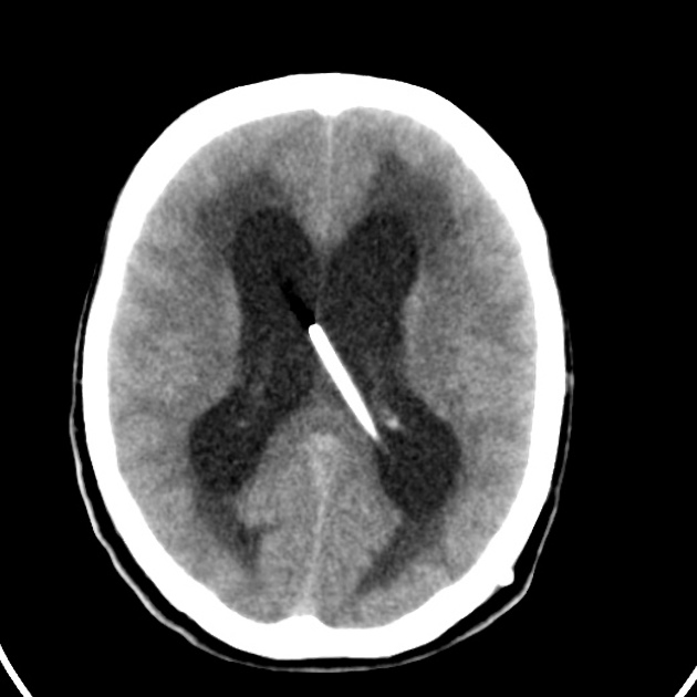
CT
low attenuation periventricular changes around the lateral ventricles
effacement of adjacent cerebral sulci may be seen, which is helpful to distinguish the condition from age-related cerebral atrophy with small vessel periventricular ischaemic disease
other corresponding features of obstructive hydrocephalus may be noted
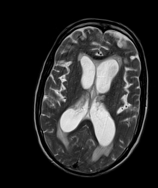
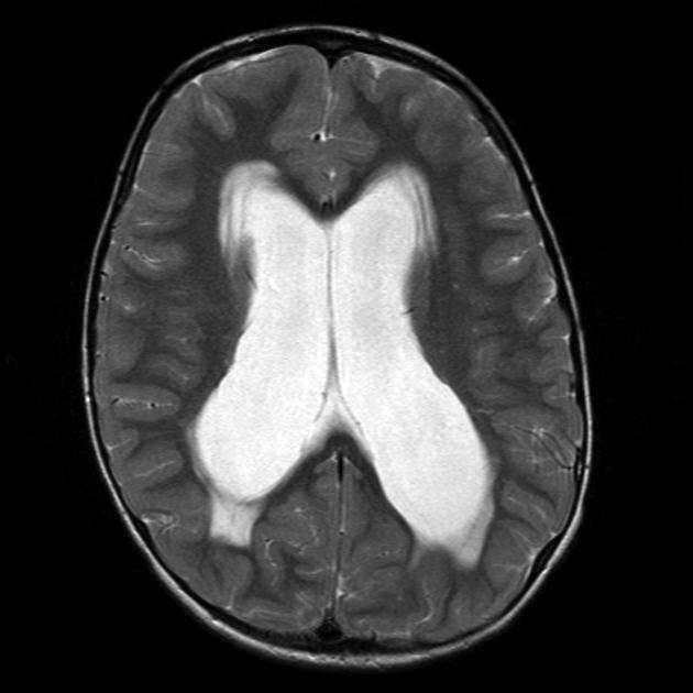
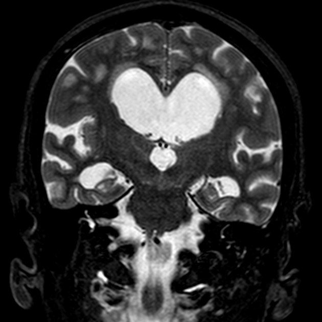
MRI
halo of high T2 or FLAIR signal around the lateral ventricles
Practical points
It is important to distinguish interstitial oedema from a commonly seen variant consisting of a slight increase in signal anterior to the frontal horns and posterior to the occipital horns, known as ependymitis granularis 3.


 Unable to process the form. Check for errors and try again.